. 2009 Nov 1;4(6):761-773.
doi: 10.2217/fnl.09.54.

Is abnormal axonal transport a cause, a contributing factor or a consequence of the neuronal pathology in Alzheimer's disease?

Affiliations

Is abnormal axonal transport a cause, a contributing factor or a consequence of the neuronal pathology in Alzheimer's disease?

Virgil Muresan et al. Future Neurol. .

Abstract

Axonal transport, the process by which membrane-bound organelles and soluble protein complexes are transported into and out of axons, ensures proper function of the neuron, including that of the synapse. As such, abnormalities in axonal transport could lead to neuronal pathology and disease. Similar to many neurodegenerative diseases, axonal transport is deficient in Alzheimer's disease (AD), a neurodegenerative brain disorder that affects old-age humans and is characterized by the deterioration of cognitive function and progressive memory loss. It was proposed that the synaptic pathology and neuronal degeneration that develops in AD could be caused by an abnormal axonal transport, and that the mutated proteins that cause early-onset AD, as well as the genetic variants that confer predisposition to late-onset AD might somehow impede axonal transport. This paper analyzes the data that support or contradict this hypothesis. Together, they indicate that, although abnormalities in axonal transport are part of the disease, additional studies are required to clearly establish to what extent deficient axonal transport is the cause or the effect of the neuronal pathology in AD, and to identify mechanisms that lead to its perturbation.


Figures

Figure 1
Figure 1. Processing of amyloid-β precursor protein via the β-secretase pathway
The positions of the transmembrane domain, of Aβ (red), and of the cleavage sites are marked. The APP-derived proteolytic fragments, sAPPβ, Aβ, AICD, and the intermediary cleavage product, CTF-β, are also shown. The cleavage by γ-secretase at positions 636/638 generates Aβ40/42. Aβ: Amyloidβ; APP: Amyloid-β precursor protein; AICD: APP intracellular domain; CTF-β: Carboxy-terminal fragment-β. Modified from [4,61].
Figure 2
Figure 2. Amyloid-β precursor protein recruits kinesin motors to cargo vesicles via adaptor proteins
JIP-1, Fe65 and Mint1/X11α bind to APP via their phosphotyrosin-binding domains. JIP-1 and Fe65 also bind to the light chains of kinesin-1; Mint1/X11α binds to KIF17, a kinesin-2 motor with dendritic targeting. In the cellular context, JIP-1 binds preferentially to phosphorylated APP, while Fe65 binds to nonphosphorylated APP. The direct binding of kinesin-1 to APP, as shown to the left, could be less relevant for the in vivo situation. APP: Amyloid-β precursor protein; JIP: JNK-interacting protein.
Figure 3
Figure 3. Hypothetical models for the release of kinesin-1 from transport vesicles, leading to disruption of the fast axonal transport in Alzheimer’s disease
(A) A slowed down axonal transport at old age could allow premature processing of APP and release of kinesin-1 from the transport vesicle, leading to a halt in transport (crossed arrow) [26]. The cessation of transport further facilitates cleavage of APP molecules by cotransported secretases (not depicted), thus amplifying the loop where the deficiencies in axonal transport and processing of APP potentiate each other. Although this model is no longer regarded as a major mechanism operating in Alzheimer’s disease (AD), the basic principle for the disruption of the axonal transport by premature release of the motor from the transport vesicle remains valid. (B) The regulation of axonal transport by phosphorylation of the light chains of kinesin-1 by casein kinase 2 (CK2) [69] or glycogen synthase kinase 3β (GSK3β) [77,78]. The phosphorylated kinesin-1 is released from the vesicle, causing a halt in transport (crossed arrow). The signaling cascades leading to phosphorylation could be triggered by the AD-linked increased production of Aβ oligomers, or by a mutated presenilin-1 (PS1swe) present in some cases of early-onset AD. It should be noted that, (A) kinesin-1 binds to APP or (B) to another receptor protein. Other cargo proteins are shown. Aβ: Amyloid-β; APP: Amyloid-β precursor protein.
Figure 4
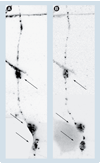
Figure 4. Clusters of mitochondria colocalize with amyloid-β deposits
Clusters of mitochondria colocalize with amyloid-β (Aβ) deposits (arrows) within the neurite of a CAD cell (a CNS-derived neuronal cell line [117]). Neurites containing Aβ deposits also contain fewer mitochondria [73]. (A) The mitochondria and (B) Aβ accumulations were detected with an antibody to lipoic acid and antibody 6E10 (Signet, Dedham, MA, USA), respectively. The inverted, fluorescence microscopy images were adjusted for contrast and brightness, to facilitate the evaluation of colocalization.
Figure 5
Figure 5. Relationship between abnormal axonal transport and axonal pathology in Alzheimer’s disease
(A) Deficiencies in axonal transport (caused by factors that normally appear at old age or by mutated proteins with a role in axonal transport) inflict axonal pathology by depriving the axon of essential factors, and/or by causing an abnormal/increased production of toxic amyloid-β (Aβ). In this case, the axonal transport deficiency is the cause of the axonal pathology. (B) An initial event, not related to axonal transport, induces abnormal/increased production of toxic Aβ, which blocks axonal transport and leads to axonal pathology. Although here, the axonal transport is not itself the initial cause, it is an essential part of the mechanism that leads to axonal pathology. (C) Axonal pathology is caused by a toxic effect of Aβ on the synaptic function, independently of axonal transport (see [31] for example), triggering neuronal death by a dying back mechanism [118], when axonal transport is also perturbed. In this case, the deficiencies in axonal transport are a result of the neuronal pathology of Alzheimer’s disease. The mechanisms described above are focused on Aβ. Other scenarios, where the abnormalities in axonal transport are a cause, a facilitating factor or a result of the neuronal pathology can be envisioned. Aβ: Amyloid-β.

Similar articles

Cited by

References

    1. Goedert M, Spillantini MG. A century of Alzheimer’s disease. Science. 2006;314:777–781. - PubMed
    1. Selkoe DJ. Alzheimer’s disease: genes, proteins, and therapy. Physiol. Rev. 2001;81:741–766. - PubMed
    1. Sisodia SS. Alzheimer’s disease: perspectives for the new millennium. J. Clin. Invest. 1999;104:1169–1170. - PMC - PubMed
    1. Selkoe DJ. Translating cell biology into therapeutic advances in Alzheimer’s disease. Nature. 1999;399:A23–A31. - PubMed
    1. Hedera P, Turner RS. Inherited dementias. Neurol. Clin. 2002;20:779–808. vii. - PubMed

LinkOut - more resources