

Upregulation of defensins in burn sheep small intestine
- PMID: 20076788
- PMCID: PMC2803769
Upregulation of defensins in burn sheep small intestine
Abstract
Objective: The aim of this study was to visualize and localize the sheep antimicrobials, beta-defensins 1, 2, and 3, (SBD-1, SBD-2, SBD-3), sheep neutrophil defensin alpha (SNP-1), and the cathelicidin LL-37 in sheep small intestine after burn injury, our hypothesis being that these compounds would be upregulated in an effort to overcome a compromised endothelial lining. Response to burn injury includes the release of proinflammatory cytokines and systemic immune suppression that, if untreated, can progress to multiple organ failure and death, so protective mechanisms have to be initiated and implemented.
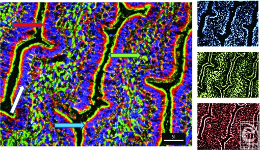
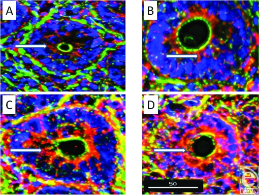
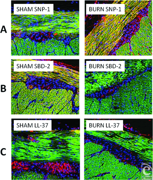
Methods: Tissue sections were probed with antibodies to the antimicrobials and then visualized with fluorescently labeled secondary antibodies and subjected to fluorescence deconvolution microscopy and image reconstruction.
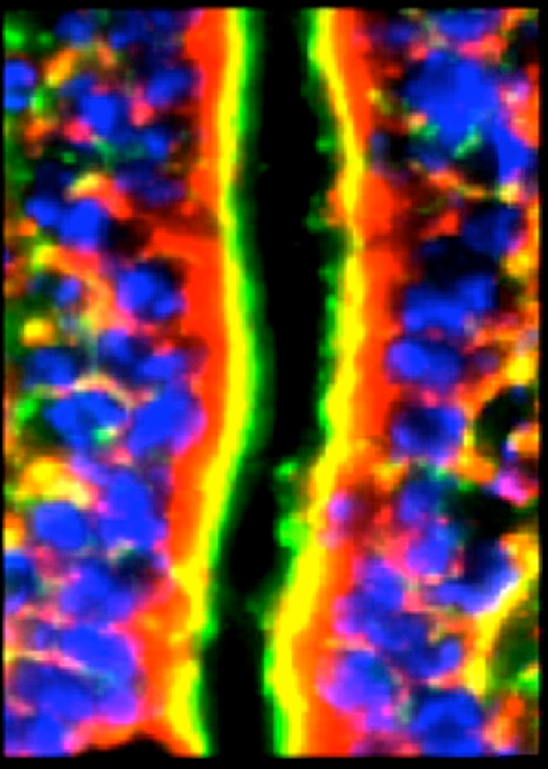
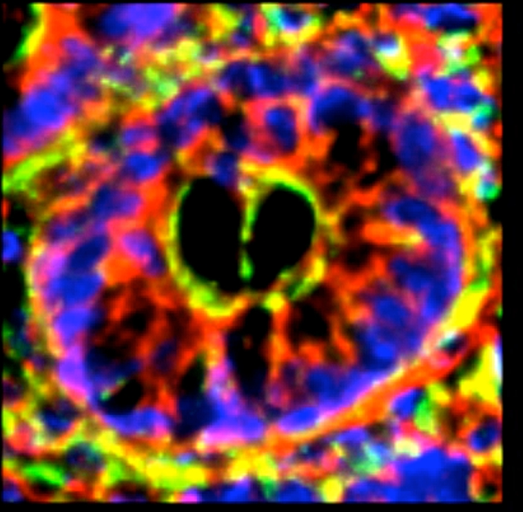
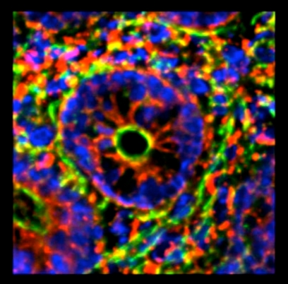
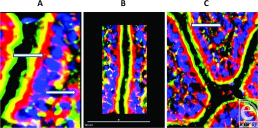
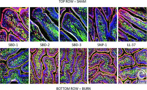
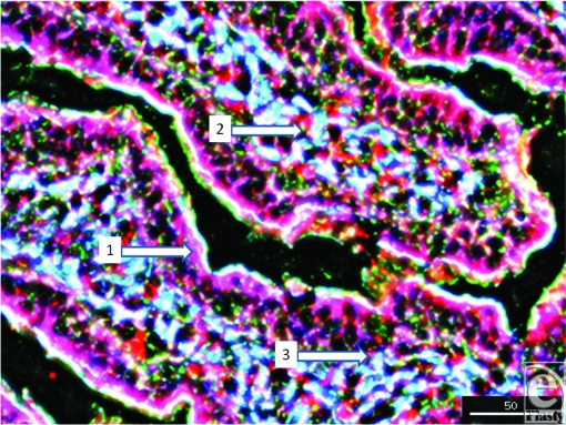
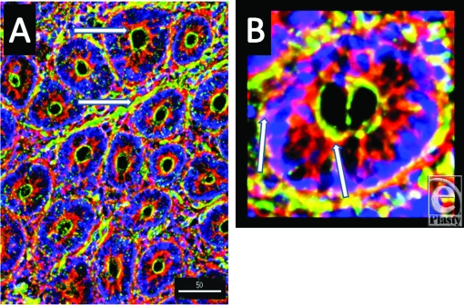
Results: In both the sham and burn samples, all the aforementioned antimicrobials were seen in each of the layers of small intestine, the highest concentration being localized to the epithelium. SBD-2, SBD-3, and SNP-1 were upregulated in both enterocytes and Paneth cells, while SNP-1 and LL-37 showed increases in both the inner circular and outer longitudinal muscle layers of the muscularis externa following burn injury. Each of the defensins, except SBD-1, was also seen in between the muscle layers of the externa and while burn caused slight increases of SBD-2, SBD-3, and SNP-1 in this location, LL-37 content was significantly decreased.
Conclusion: That while each of these human antimicrobials is present in multiple layers of sheep small intestine, SBD-2, SBD-3, SNP-1, and LL-37 are upregulated in the specific layers of the small intestine.
Figures

Similar articles
-
Alterations in content and localization of defensins in rat ileum and jejunum following ischemia-reperfusion. Specific peptides, in specific places, for specific jobs?Eplasty. 2011 Feb 23;11:e8. Eplasty. 2011. PMID: 21369366 Free PMC article.
-
Immunofluorescence deconvolution microscopy and image reconstruction of human defensins in normal and burned skin.J Burns Wounds. 2005 Apr 25;4:e7. J Burns Wounds. 2005. PMID: 16921412 Free PMC article.
-
Developmental expression and distribution of sheep beta-defensin-2.Dev Comp Immunol. 2004 Feb;28(2):171-8. doi: 10.1016/s0145-305x(03)00105-8. Dev Comp Immunol. 2004. PMID: 12969802
-
Defensin deficiency, intestinal microbes, and the clinical phenotypes of Crohn's disease.J Leukoc Biol. 2005 Apr;77(4):460-5. doi: 10.1189/jlb.0904543. Epub 2004 Dec 23. J Leukoc Biol. 2005. PMID: 15618294 Review.
-
Defensin-mediated innate immunity in the small intestine.Best Pract Res Clin Gastroenterol. 2004 Apr;18(2):405-19. doi: 10.1016/j.bpg.2003.10.010. Best Pract Res Clin Gastroenterol. 2004. PMID: 15123078 Review.
Cited by
-
Characterization of paneth cells in alpacas (Vicugna pacos, Mammalia, Camelidae).Tissue Cell. 2016 Aug;48(4):383-8. doi: 10.1016/j.tice.2016.04.003. Epub 2016 May 7. Tissue Cell. 2016. PMID: 27233914 Free PMC article.
-
The Paneth Cell: The Curator and Defender of the Immature Small Intestine.Front Immunol. 2020 Apr 3;11:587. doi: 10.3389/fimmu.2020.00587. eCollection 2020. Front Immunol. 2020. PMID: 32308658 Free PMC article. Review.
-
Alterations in content and localization of defensins in rat ileum and jejunum following ischemia-reperfusion. Specific peptides, in specific places, for specific jobs?Eplasty. 2011 Feb 23;11:e8. Eplasty. 2011. PMID: 21369366 Free PMC article.
References
-
- Sheridan RL, Tompkins RG. Etiology and prevention of multiple organ failure. In: Herndon D, editor. Total Burn Care. 2nd ed. Philadelphia: Saunders; 2001. p. 382.
-
- Schwacha MG. Macrophages and post-burn immune dysfunction. Burns. 2003;29:1–14. - PubMed
-
- Bick RJ, Poindexter BJ, Bhat S, Gulati S, Buja M, Milner SM. Effects of cytokines and heat shock on defensin levels of cultured keratinocytes. Burns. 2004;30:329–33. - PubMed
-
- Milner SM, Ortega MR. Reduced antimicrobial peptide expression in human burn wounds. Burns. 1999;25:411–3. - PubMed
-
- Ortega MR, Ganz T, Milner SM. Human beta defensin in burn blister fluid. Burns. 2000;26:724–6. - PubMed
LinkOut - more resources
Full Text Sources