Review
. 2010 May;15(5):527-40.
doi: 10.1007/s10495-009-0452-5.

Regulation of apoptosis-associated lysosomal membrane permeabilization

Affiliations
Review

Regulation of apoptosis-associated lysosomal membrane permeabilization

Ann-Charlotte Johansson et al. Apoptosis. 2010 May.

Abstract

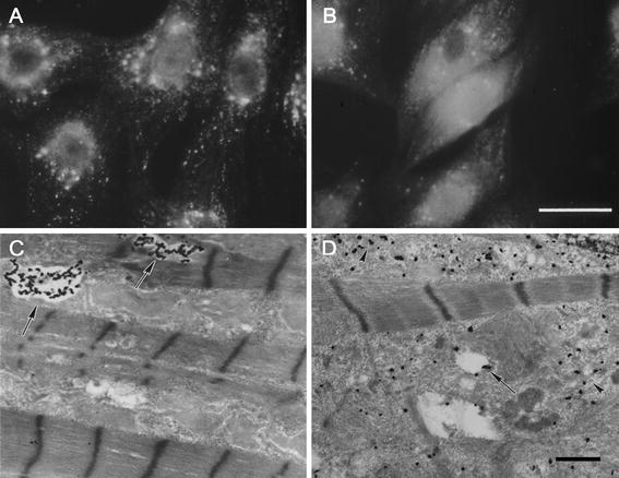
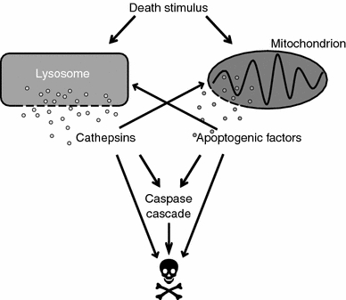
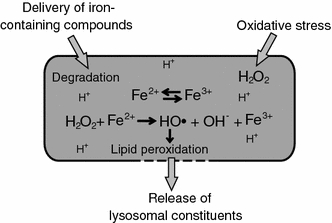
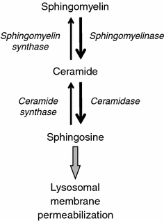
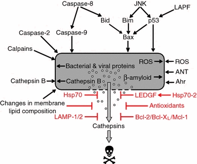
Lysosomal membrane permeabilization (LMP) occurs in response to a large variety of cell death stimuli causing release of cathepsins from the lysosomal lumen into the cytosol where they participate in apoptosis signaling. In some settings, apoptosis induction is dependent on an early release of cathepsins, while under other circumstances LMP occurs late in the cell death process and contributes to amplification of the death signal. The mechanism underlying LMP is still incompletely understood; however, a growing body of evidence suggests that LMP may be governed by several distinct mechanisms that are likely engaged in a death stimulus- and cell-type-dependent fashion. In this review, factors contributing to permeabilization of the lysosomal membrane including reactive oxygen species, lysosomal membrane lipid composition, proteases, p53, and Bcl-2 family proteins, are described. Potential mechanisms to safeguard lysosomal integrity and confer resistance to lysosome-dependent cell death are also discussed.


Figures

Fig. 1
Fig. 1
Apoptosis-associated release of lysosomal cathepsin D into the cytosol. Visualization of cathepsin D in rat cardiomyocytes exposed to the redox-cycling drug naphthazarin by immunofluorescence microscopy (a and b) or immuno-electron microscopy using antibodies tagged with ultra-small gold particles and subsequent silver enhancement (c and d). There is a shift from a punctate lysosomal staining pattern in control cells (a) to a diffuse cytosolic staining in cells exposed to naphthazarin (b). Likewise, in electron micrographs (c), cathepsin D can be seen in lysosome-like structures in control cells (arrows), while in cells treated with naphthazarin (d), cathepsin D is spread throughout the cytosol (arrow heads), bars = 30 μm (a and b) and 1 μm (c and d)
Fig. 2
Fig. 2
Lysosomal participation in cell death signaling. Lysosomal membrane permeabilization (LMP), i.e., release of cathepsins from lysosomes into the cytosol, is an important step in cell death signaling induced by a variety of stimuli, such as death receptor activation, radiation, and cytotoxic drugs. Several cytosolic cathepsin targets have been described and the signaling downstream LMP involves both caspase-dependent and -independent pathways. Engagement of the mitochondrial pathway is a common downstream event of LMP, but cathepsins may also cause cell death without involvement of mitochondria
Fig. 3
Fig. 3
Destabilization of the lysosomal membrane by lipid peroxidation. In response to oxidative stress, increased amounts of hydrogen peroxide can diffuse into the lysosome. In the lysosomal lumen, the acidic milieu and the presence of low-molecular-weight iron, derived from degraded iron-containing proteins, promotes the reduction of iron and the generation of hydroxyl radicals. The hydroxyl radicals induce peroxidation of membrane lipids and thereby cause leakage of lysosomal constituents into the cytosol
Fig. 4
Fig. 4
Accumulation of sphingosine leads to lysosomal membrane permeabilization. An apoptotic stimulus, e.g., tumor necrosis factor-α, leads to increased activity of sphingomyelinase, which catalyzes the formation of ceramide from sphingomyelin. By the action of ceramidase, ceramide is then converted into sphingosine, which accumulates in lysosomes and acquires detergent-like properties, leading to lysosomal membrane permeabilization
Fig. 5
Fig. 5
Factors regulating lysosomal membrane permeabilization. This schematic presents a number of factors that may be responsible for lysosomal membrane permeabilization (LMP). The relative importance of each mechanism likely depends on the cell type and death stimulus. Mechanisms that are believed to safeguard lysosomal integrity and protect from lysosome-mediated cell death are also shown. “Changes in membrane lipid composition” includes membrane destabilizing factors such as phospholipase A2 and sphingosine, as well as LPM protective substances e.g., cholesterol and sphingomyelin. Abbreviations: JNK, c-Jun N-terminal kinase; LAPF, lysosome-associated apoptosis-inducing protein containing the pleckstrin homology and FYVE domains; Hsp, heat shock protein; ROS, reactive oxygen species; ANT, adenine nucleotide translocator; Ahr, aryl hydrocarbon receptor; LEDGF, lens epithelium-derived growth factor; LAMP, lysosome-associated membrane protein

References

    1. de Duve C, Pressman BC, Gianetto R, Wattiaux R, Applemans F. Tissue fractionation studies. 6. Intracellular distribution patterns of enzymes in rat-liver tissue. Biochem J. 1955;60:604–617. - PMC - PubMed
    1. de Duve C. Lysosomes revisited. Eur J Biochem. 1983;137:391–397. doi: 10.1111/j.1432-1033.1983.tb07841.x. - DOI - PubMed
    1. Turk B, Stoka V. Protease signalling in cell death: caspases versus cysteine cathepsins. FEBS Lett. 2007;581:2761–2767. doi: 10.1016/j.febslet.2007.05.038. - DOI - PubMed
    1. de Duve C. Lysosomes, a new group of cytoplasmic particles. In: Hayashi T, editor. Subcellular particles. New York: Ronald Press; 1959. pp. 128–159.
    1. Roberg K, Öllinger K. Oxidative stress causes relocation of the lysosomal enzyme cathepsin D with ensuing apoptosis in neonatal rat cardiomyocytes. Am J Pathol. 1998;152:1151–1156. - PMC - PubMed

MeSH terms