

Quantification of trabecular bone microdamage using the virtual internal bond model and the individual trabeculae segmentation technique
- PMID: 20077238
- PMCID: PMC3384516
- DOI: 10.1080/10255840903405660
Quantification of trabecular bone microdamage using the virtual internal bond model and the individual trabeculae segmentation technique
Abstract
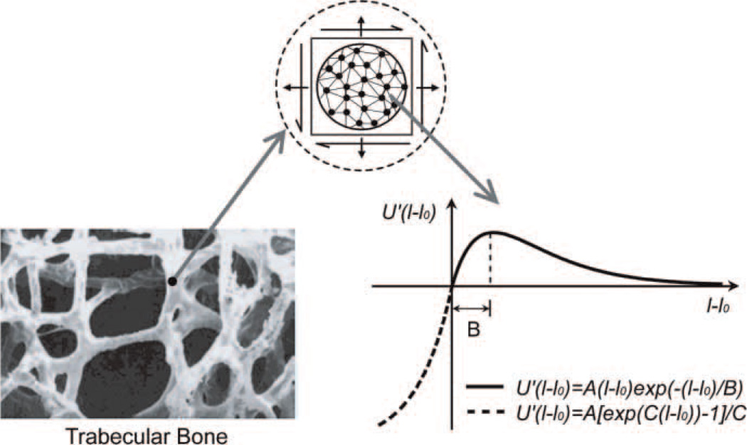
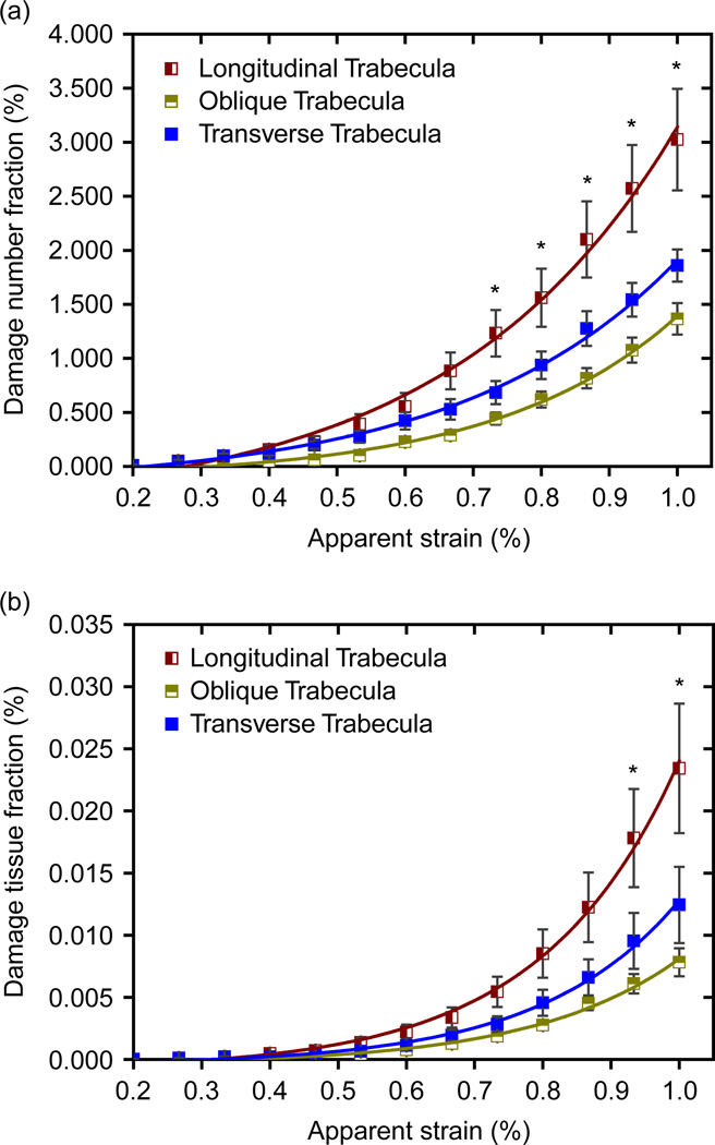
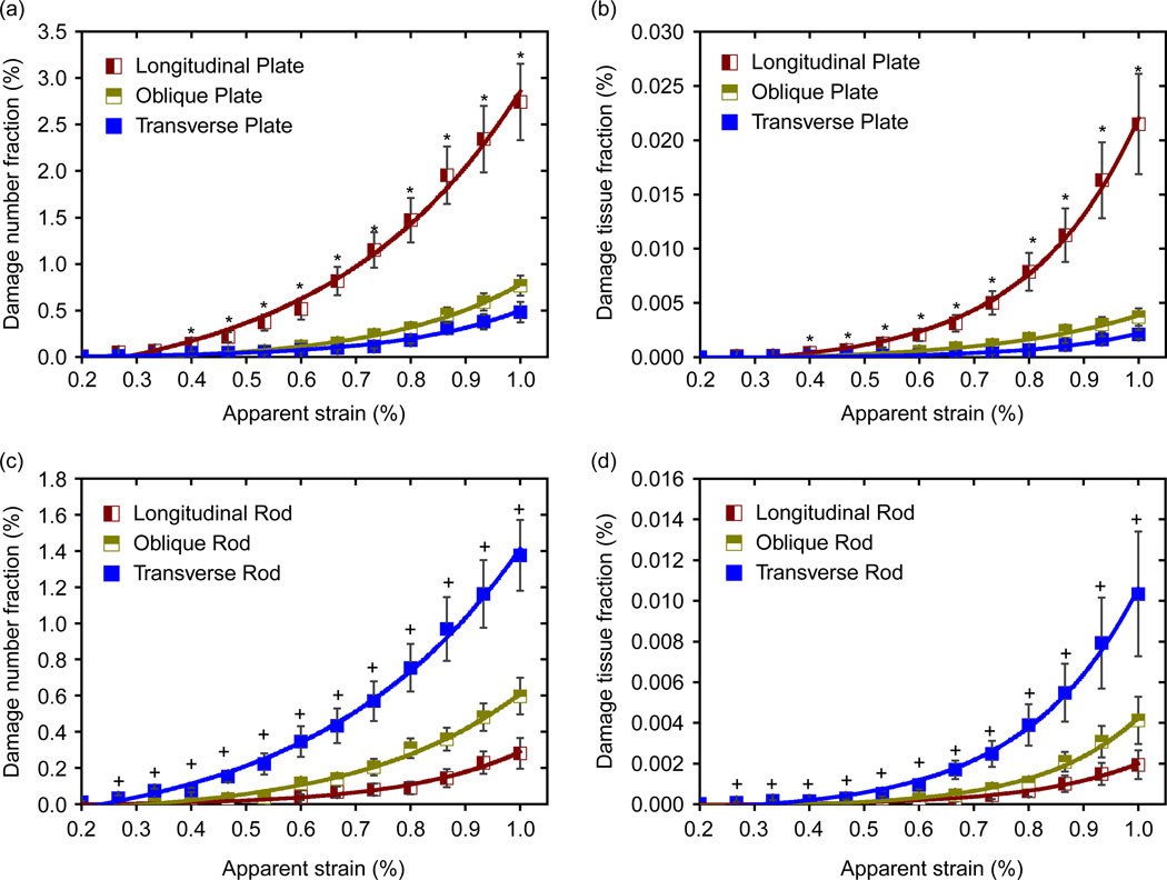
Trabecular bone microdamage significantly influences the skeletal integrity and bone remodelling process. In this paper a novel constitutive model, called the virtual internal bond model (VIB), was adopted for simulating the damage behaviour of bone tissue. A unique 3D image analysis technique, named individual trabeculae segmentation, was used to analyse the effects of microarchitectures on the damage behaviours of trabecular bone. We demonstrated that the process of initiation and accumulation of microdamage in trabecular bone samples can be captured by the VIB-embedded finite-element method simulation without a separate fracture criterion. Our simulation results showed that the microdamage can occur at as early as about 0.2-0.4% apparent strain, and a large volume of microdamage was accumulated around the apparent yield strain. In addition we found that the plate-like trabeculae, especially the longitudinal ones, take crucial roles in the microdamage behaviours of trabecular bone.
Conflict of interest statement
The authors state that they have no any financial, professional and personal conflict of interests.
Figures


Similar articles
-
Relative roles of microdamage and microfracture in the mechanical behavior of trabecular bone.J Orthop Res. 2001 Nov;19(6):1001-7. doi: 10.1016/S0736-0266(01)00053-5. J Orthop Res. 2001. PMID: 11780997
-
Age-related changes in human trabecular bone: Relationship between microstructural stress and strain and damage morphology.J Biomech. 2011 Aug 11;44(12):2279-85. doi: 10.1016/j.jbiomech.2011.05.034. Epub 2011 Jul 2. J Biomech. 2011. PMID: 21724189 Free PMC article.
-
Predicting trabecular bone microdamage initiation and accumulation using a non-linear perfect damage model.Med Eng Phys. 2008 Jul;30(6):725-32. doi: 10.1016/j.medengphy.2007.02.011. Epub 2007 Sep 18. Med Eng Phys. 2008. PMID: 17881275
-
The structural and biomechanical basis of the gain and loss of bone strength in women and men.Endocrinol Metab Clin North Am. 2003 Mar;32(1):25-38. doi: 10.1016/s0889-8529(02)00078-6. Endocrinol Metab Clin North Am. 2003. PMID: 12699291 Review.
-
[Bone fracture and the healing mechanisms. Microdamage and Microfracture].Clin Calcium. 2009 May;19(5):699-703. Clin Calcium. 2009. PMID: 19398838 Review. Japanese.
Cited by
-
Determinants of bone damage: An ex-vivo study on porcine vertebrae.PLoS One. 2018 Aug 16;13(8):e0202210. doi: 10.1371/journal.pone.0202210. eCollection 2018. PLoS One. 2018. PMID: 30114229 Free PMC article.
-
A coupled mechano-biochemical model for bone adaptation.J Math Biol. 2014 Dec;69(6-7):1383-429. doi: 10.1007/s00285-013-0736-9. Epub 2013 Nov 12. J Math Biol. 2014. PMID: 24212399
-
Can deterministic mechanical size effects contribute to fracture and microdamage accumulation in trabecular bone?J Theor Biol. 2010 Jul 21;265(2):202-10. doi: 10.1016/j.jtbi.2010.04.009. Epub 2010 Apr 14. J Theor Biol. 2010. PMID: 20398678 Free PMC article.
-
Distributions of Microdamage Are Altered Between Trabecular Rods and Plates in Cancellous Bone From Men With Type 2 Diabetes Mellitus.J Bone Miner Res. 2022 Apr;37(4):740-752. doi: 10.1002/jbmr.4509. Epub 2022 Feb 15. J Bone Miner Res. 2022. PMID: 35064941 Free PMC article.
-
Bone-inspired microarchitectures achieve enhanced fatigue life.Proc Natl Acad Sci U S A. 2019 Dec 3;116(49):24457-24462. doi: 10.1073/pnas.1905814116. Epub 2019 Nov 18. Proc Natl Acad Sci U S A. 2019. PMID: 31740616 Free PMC article.
References
-
- Bevill G, Eswaran SK, Gupta A, Papadopoulos P, Keaveny TM. Influence of bone volume fraction and architecture on computed large-deformation failure mechanisms in human trabecular bone. Bone. 2006;39(6):1218–1225. - PubMed
-
- Borah B, Dufresne TE, Cockman MD, Gross GJ, Sod EW, Myers WR, Combs KS, Higgins RE, Pierce SA, Stevens ML. Evaluation of changes in trabecular bone architecture and mechanical properties of minipig vertebrae by three-dimensional magnetic resonance microimaging and finite element modeling. J Bone Miner Res. 2000;15(9):1786–1797. - PubMed
-
- Burr D. Microdamage and bone strength. Osteoporos Int. 2003:S67–S72. - PubMed
-
- Burr DB, Forwood MR, Fyhrie DP, Martin B, Schaffler MB, Turner CH. Bone microdamage acid skeletal fragility in osteoporotic and stress fractures. J Bone Miner Res. 1997;12(1):6–15. - PubMed
-
- Charras GT, Guldberg RE. Improving the local solution accuracy of large-scale digital image-based finite element analyses. J Biomech. 2000;33(2):255–259. - PubMed
Publication types
MeSH terms
Grants and funding
LinkOut - more resources
Full Text Sources