

Kinematic and electromyographic tools for characterizing movement disorders in mice
- PMID: 20077474
- PMCID: PMC2925152
- DOI: 10.1002/mds.22933
Kinematic and electromyographic tools for characterizing movement disorders in mice
Abstract
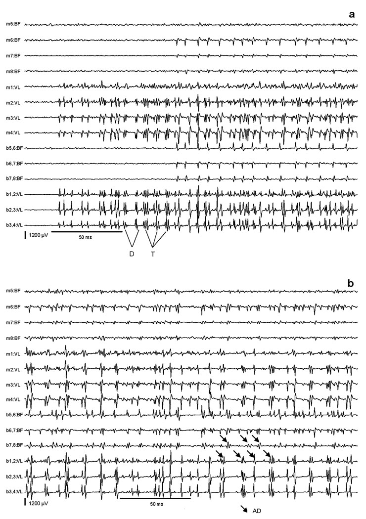
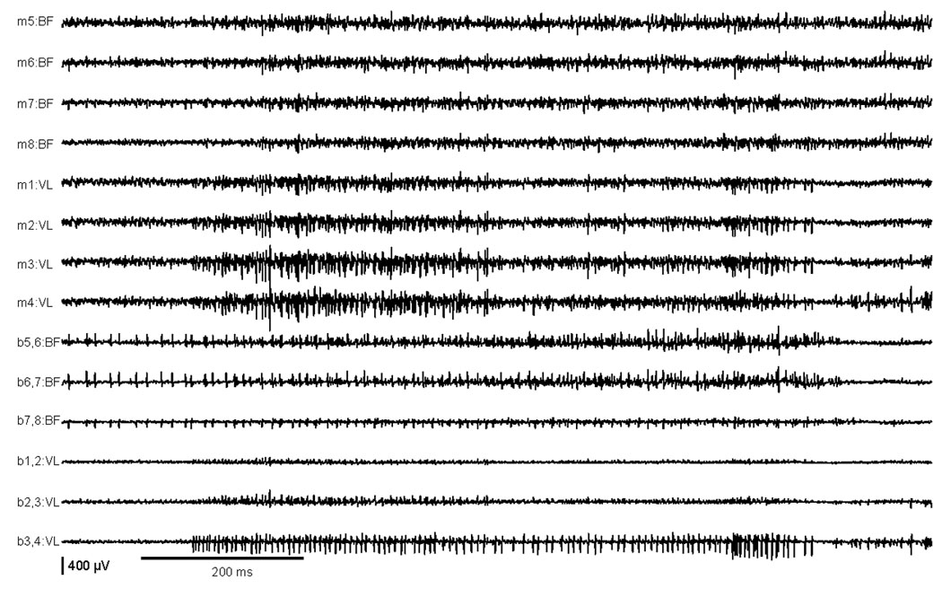
Increasing interest in rodent models for movement disorders has led to an increasing need for more accurate and precise methods for both delineating the nature of abnormal movements and measuring their severity. These studies describe application of simultaneous high-speed video kinematics with multichannel electromyography (EMG) to characterize the movement disorder exhibited by tottering mutant mice. These mice provide a uniquely valuable model, because they exhibit paroxysmal dystonia superimposed on mild baseline ataxia, permitting the examination of these two different problems within the same animals. At baseline with mild ataxia, the mutants exhibited poorly coordinated movements with increased variation of stance and swing times, and slower spontaneous walking velocities. The corresponding EMG showed reduced mean amplitudes of biceps femoris and vastus lateralis, and poorly modulated EMG activities during the step cycle. Attacks of paroxysmal dystonia were preceded by trains of EMG bursts with doublets and triplets simultaneously in the biceps femoris and vastus lateralis followed by more sustained coactivation. These EMG characteristics are consistent with the clinical phenomenology of the motor phenotype of tottering mice as a baseline of mild ataxia with intermittent attacks of paroxysmal dystonia. The EMG characteristics of ataxia and dystonia in the tottering mice also are consistent with EMG studies of other ataxic or dystonic animals and humans. These studies provide insights into how these methods can be used for delineating movement disorders in mice and for how they may be compared with similar disorders of humans.
(c) 2010 Movement Disorder Society.
Figures
Similar articles
-
Potassium channel blockers inhibit the triggers of attacks in the calcium channel mouse mutant tottering.J Neurosci. 2005 Apr 20;25(16):4141-5. doi: 10.1523/JNEUROSCI.0098-05.2005. J Neurosci. 2005. PMID: 15843617 Free PMC article.
-
Low-frequency oscillations in the cerebellar cortex of the tottering mouse.J Neurophysiol. 2009 Jan;101(1):234-45. doi: 10.1152/jn.90829.2008. Epub 2008 Nov 5. J Neurophysiol. 2009. PMID: 18987121 Free PMC article.
-
Kinematic and electromyographic analysis in patients with patellofemoral pain syndrome during single leg triple hop test.Gait Posture. 2016 Sep;49:246-251. doi: 10.1016/j.gaitpost.2016.07.020. Epub 2016 Jul 19. Gait Posture. 2016. PMID: 27470227
-
[Assessment of motor impairment with electromyography--the kinesiological EMG].Ideggyogy Sz. 2003 Nov 20;56(11-12):360-9. Ideggyogy Sz. 2003. PMID: 14743590 Review. Hungarian.
-
Contributions to the understanding of gait control.Dan Med J. 2014 Apr;61(4):B4823. Dan Med J. 2014. PMID: 24814597 Review.
Cited by
-
Electromyographic evidence in support of a knock-in mouse model of DYT1 Dystonia.Mov Disord. 2016 Nov;31(11):1633-1639. doi: 10.1002/mds.26677. Epub 2016 May 31. Mov Disord. 2016. PMID: 27241685 Free PMC article.
-
Altered brain state during episodic dystonia in tottering mice decouples primary motor cortex from limb kinematics.Dystonia. 2023;2:10974. doi: 10.3389/dyst.2023.10974. Epub 2023 Feb 2. Dystonia. 2023. PMID: 37800168 Free PMC article.
-
Quantifying muscle alterations in a Parkinson's disease animal model using electromyographic biomarkers.Med Biol Eng Comput. 2021 Sep;59(9):1735-1749. doi: 10.1007/s11517-021-02400-3. Epub 2021 Jul 23. Med Biol Eng Comput. 2021. PMID: 34297299
-
Limited regional cerebellar dysfunction induces focal dystonia in mice.Neurobiol Dis. 2013 Jan;49:200-10. doi: 10.1016/j.nbd.2012.07.019. Epub 2012 Jul 28. Neurobiol Dis. 2013. PMID: 22850483 Free PMC article.
-
Deep Brain Stimulation of the Interposed Nucleus Reverses Motor Deficits and Stimulates Production of Anti-inflammatory Cytokines in Ataxia Mice.Mol Neurobiol. 2022 Jul;59(7):4578-4592. doi: 10.1007/s12035-022-02872-w. Epub 2022 May 17. Mol Neurobiol. 2022. PMID: 35581519
References
-
- Jinnah HA, Hess EJ. The assessment of movement disorders in mice. In: Le Doux MS, editor. Animal models of movement disorders. San Diego: Elsevier Academic Press; 2005. pp. 55–71.
-
- Carter GT, Longley KJ, Entrikin RK. Electromyographic and nerve conduction studies in the mdx mouse. Am J Phys Med Rehabil. 1992 Feb. 571 - PubMed
-
- Chandran AP, Oda K, Shibasaki H, Kikuchi T, Pisharodi M. Stimulus induced repetitive muscle potentials in the gracile axonal dystrophy (GAD) mouse. Electromygr Clinc Neurophysiol. 1995;35:225–230. - PubMed
-
- Entrikin RK, Abresch RT, Sharman RB, Larson DB, Levine NA. Contractile and EMG studies of murine myotonia (mto) and muscular dystrophy (dy/dy) Muscle Nerve. 1987;10:293–298. - PubMed
-
- Shirakawa T, Sakai K, Kitagawa Y, Hori A, Hirose G. A novel murine myotonia congenita without molecular defects in the CLC-1 and the SCN4A. Neurology. 2002;59:1091–1094. - PubMed
Publication types
MeSH terms
Substances
Grants and funding
LinkOut - more resources
Full Text Sources
Medical
