Review
. 2010 Sep-Oct;29(5):830-44.
doi: 10.1002/mas.20269.

Glycoprotein analysis using protein microarrays and mass spectrometry

Affiliations
Review

Glycoprotein analysis using protein microarrays and mass spectrometry

Tasneem Patwa et al. Mass Spectrom Rev. 2010 Sep-Oct.

Abstract

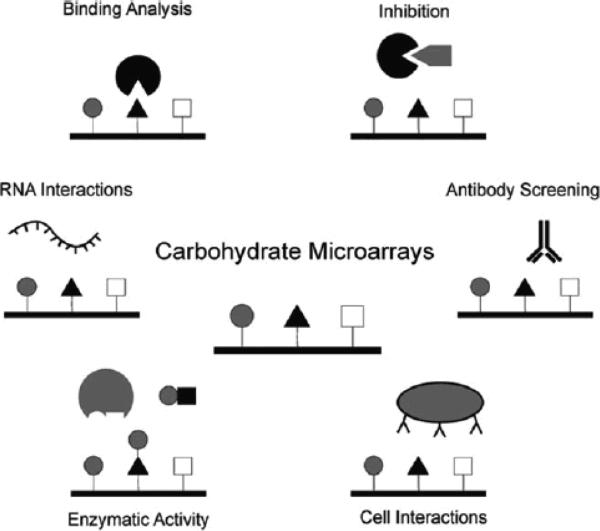
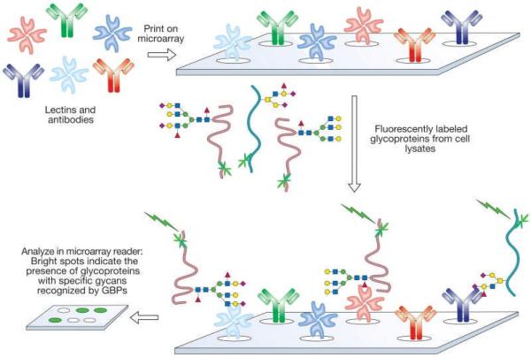
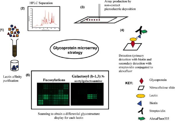
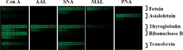
Protein glycosylation plays an important role in a multitude of biological processes such as cell-cell recognition, growth, differentiation, and cell death. It has been shown that specific glycosylation changes are key in disease progression and can have diagnostic value for a variety of disease types such as cancer and inflammation. The complexity of carbohydrate structures and their derivatives makes their study a real challenge. Improving the isolation, separation, and characterization of carbohydrates and their glycoproteins is a subject of increasing scientific interest. With the development of new stationary phases and molecules that have affinity properties for glycoproteins, the isolation and separation of these compounds have advanced significantly. In addition to detection with mass spectrometry, the microarray platform has become an essential tool to characterize glycan structure and to study glycosylation-related biological interactions, by using probes as a means to interrogate the spotted or captured glycosylated molecules on the arrays. Furthermore, the high-throughput and reproducible nature of microarray platforms have been highlighted by its extensive applications in the field of biomarker validation, where a large number of samples must be analyzed multiple times. This review covers a brief survey of the other experimental methodologies that are currently being developed and used to study glycosylation and emphasizes methodologies that involve the use of microarray platforms. This review describes recent advances in several options of microarray platforms used in glycoprotein analysis, including glycoprotein arrays, glycan arrays, lectin arrays, and antibody/lectin arrays. The translational use of these arrays in applications related to characterization of cells and biomarker discovery is also included.


Figures

Figure 1
Figure 1
Application range of carbohydrate-microarray experiments. The carbohydrate-ligand specificity for carbohydrate binding molecules has been assessed. Screening for inhibitors of carbohydrate-mediated interactions and determination of IC50 values can be performed by a co-incubation of the binding molecule with an inhibitor. Sugar interactions of an entire organism such as a whole cell or virus can be determined without purifying the carbohydrate-binding proteins. Reprinted and adapted with permission from Horlacher & Seeberger (2008). Copyright 2008, Royal Society Chemistry.
Figure 2
Figure 2
Analysis of cellular glycomes, using lectin and antibody arrays. The arrays were generated by immobilization of lectins and glycan-specific antibodies on a chip. Glycoproteins from cell or tissue samples were labeled with a fluorescent dye and incubated with the array. The fluorescent spots reflect the presence of glycoproteins that bear glycans recognized by the corresponding lectin or antibody. The technique provides minimal structural detail, but permits rapid high-throughput analysis of many samples. Intact cells or virus particles can also be interrogated on lectin microarrays. Reprinted and adapted with permission from Bertozzi & Sasisekharan (2009). Copyright ©2009 by The Consortium of Glycobiology Editors, La Jolla, California.
Figure 3
Figure 3
Proposed experimental strategy to study serum glycoproteins. (1) Lectin purification with a general lectin column. (2) Nonporous reversed-phase HPLC separation and fraction collection. (3) Microarray production with a noncontact piezoelectric printing device. (4) Glycan detection with biotinylated lectin-streptavidin-Alexafluor555. (5) Image acquisition and spot analysis with Genepix 6.0 software. Reprinted with permission from Patwa et al. (2006). Copyright 2006, American Chemical Society.
Figure 4
Figure 4
Scanned images of printed standard glycoproteins probed with different lectins. Each block bracketed on the right represents a dilution series of standards from 0.025 to 0.5 mg/mL. Each dilution has been printed as nine replicates in a 3X3 block. Reprinted with permission from Patwa et al. (2006). Copyright 2006, American Chemical Society.
Figure 5
Figure 5
Detection of glycans on antibody arrays. The first step of this new method is the chemical derivatization of the glycans on the spotted antibodies to block lectin binding to those glycans. The cis-hydroxyl groups of the glycans on the spotted antibodies were gently oxidized to convert them to aldehyde groups, which react with a hydrazide-maleimide bifunctional crosslinking reagent; the resulting product reacts with a Cys-Gly dipeptide. The Cys-Gly dipeptide adds bulk to the derivatized carbohydrates to hinder lectin binding. Reprinted and adapted with permission from Chen et al. (2007). Copyright 2007, Nature Publishing Group.
Figure 6
Figure 6
The MALDI-MS spectra generated from the microarray spots of Amyloid p component antibody after on-target digestion. The peaks identified as Amyloid p component were marked with green arrows, where the extra peaks that appear in c were marked with black arrows. a-control spot, without incubation with serum; b- incubated with10X-diluted serum; c-incubated with 2X-diluted serum. Reprinted and adapted with permission from Li et al. (2009). Copyright 2009, American Chemical Society.
Figure 7
Figure 7
Sections of glycoprotein microarray to compare one fraction from NPS-RP-HPLC across all 24 samples. Each panel is a section of identical arrays probed with lectin indicated on the left side of the panel. It was observed that this fraction contained proteins that were predominantly mannosylated and fucosylated. It was also observed that the level of glycosylation (based on raw microarray data) was higher in cancer samples compared to the controls. Reprinted with permission from Zhao et al. (2007). Copyright 2007, American Chemical Society.
Figure 8
Figure 8
The normalized glycoprotein microarray responses to lectin AAL were visualized with principal component analysis (PCA). Twenty-four serum samples (10 normal, 8 chronic pancreatitis, and 6 pancreatic cancers) were studied. The figure shows the clustering of serum samples obtained from patients with pancreatic cancer, chronic pancreatitis, or normal subjects. Reprinted with permission from Zhao et al. (2007). Copyright 2007, American Chemical Society.
Figure 9
Figure 9
Elevated fucosylation and sialylation of complement C3 (A) and histidine-rich glycoprotein (C) investigated with AAL and SNA blot analysis. The corresponding protein expression levels are shown in (B) for complement C3 and (D) for histidine-rich glycoprotein, respectively. Reprinted with permission from from Qiu et al. (2008). Copyright 2008, American Chemical Society.

Similar articles

Cited by

References

    1. Adams EW, Ratner DM, Bokesch HR, McMahon JB, O'Keefe BR, Seeberger PH. Oligosaccharide and glycoprotein Microarrays as tools in HIV glycobiology: Glycan-dependent gp120/protein interactions. Chem Biol. 2004;6:875–881. - PubMed
    1. Amon S, Zamfir AD, Rizzi A. Glycosylation analysis of glycoproteins and proteoglycans using capillary electrophoresis-mass spectrometry strategies. Electrophoresis. 2008;29:2485–2507. - PubMed
    1. Angeloni S, Ridet JL, Kusy N, Gao H, Crevoisier F, Guinchard S, Kochhar S, Sigrist H, Sprenger N. Glycoprofiling with micro-arrays of glycoconjugates and lectins. Glycobiology. 2005;15:31–41. - PubMed
    1. Arnold JN, Saldova R, Hamid UMA, Rudd PM. Evaluation of the serum N-linked glycome for the diagnosis of cancer and chronic inflammation. Proteomics. 2008;8:3284–3293. - PubMed
    1. Bertozzi CR, Sasisekharan R. Glycomics. In: Varki A, editor. Essentials of Glycobiology. Second Edition. The Consortium of Glycobiology Editors; California: 2009. chapter 48.

Publication types