Review
. 2010:5:297-348.
doi: 10.1146/annurev.pathol.4.110807.092314.

Mitochondrial energetics and therapeutics

Affiliations
Review

Mitochondrial energetics and therapeutics

Douglas C Wallace et al. Annu Rev Pathol. 2010.

Abstract

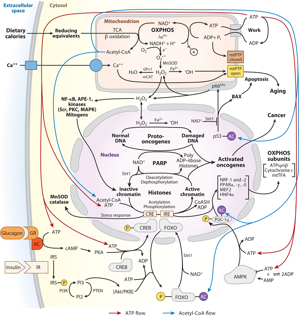
Mitochondrial dysfunction has been linked to a wide range of degenerative and metabolic diseases, cancer, and aging. All these clinical manifestations arise from the central role of bioenergetics in cell biology. Although genetic therapies are maturing as the rules of bioenergetic genetics are clarified, metabolic therapies have been ineffectual. This failure results from our limited appreciation of the role of bioenergetics as the interface between the environment and the cell. A systems approach, which, ironically, was first successfully applied over 80 years ago with the introduction of the ketogenic diet, is required. Analysis of the many ways that a shift from carbohydrate glycolytic metabolism to fatty acid and ketone oxidative metabolism may modulate metabolism, signal transduction pathways, and the epigenome gives us an appreciation of the ketogenic diet and the potential for bioenergetic therapeutics.


Figures

Figure 1
Figure 1
Bioenergetic regulation of cellular physiology and gene expression. Dietary calories enter the cell as reducing equivalents. Carbohydrates are processed through glycolysis to generate cytosolic pyruvate and NADH (reduced nicotinamide adenine nucleotide). The pyruvate then enters the mitochondrion, is processed through pyruvate dehydrogenase, and is converted to acetyl-CoA, CO2, and NADH. Fatty acids and ketone bodies enter the mitochondrion directly, where they generate acetyl-CoA and mitochondrial NADH. NADH can be oxidized within the mitochondrion by the electron transport chain to generate an inner-membrane electrochemical gradient (ΔP = Δψ + ΔµH+). This ΔP can then be used to generate ATP by the ATP synthase. The ATP is exported to the cytosol by the adenine nucleotide translocator (ANT) to energize work. Excess mitochondrial reducing equivalents can be transferred to O2 to generate superoxide anion (O2·). O2· is converted to hydrogen peroxide (H2O2) by manganese superoxide dismutase (MnSOD). H2O2 can diffuse out of the mitochondrion into the cytosol and nucleus. Further reduction of H2O2 results in hydroxyl radical (·OH). The mitochondrial permeability transition pore (mtPTP) senses mitochondrial energy decline, reactive oxygen species (ROS) production, altered oxidation-reduction (redox) state, and increased Ca2+. When activated, it opens a channel in the inner membrane, collapses ΔP, and induces apoptosis. Carbohydrate calories, in the form of glucose, are monitored by the pancreatic islet cells. High serum glucose elicits the secretion of insulin, which binds to the insulin receptors of target cells. This activates the phosphatidylinositol 3 kinase (PI3K) pathway to activate Akt protein kinase B (PKB). Akt phosphorylates the forkhead box, subgroup O (FOXO) transcription factors, barring them from the nucleus and their binding to insulin response elements (IREs). IREs are upstream of the mitochondrial transcription factor coactivator, peroxisome proliferator–activated receptor gamma coactivator 1 alpha (PGC-1α). In the absence of FOXO binding to IRE, PGC-1α transcription is reduced and mitochondrial biogenesis and oxidative phosphorylation (OXPHOS) decline, shifting metabolism toward glycolysis. When carbohydrates are limiting, serum glucose declines; insulin secretion diminishes; and the FOXOs become dephosphorylated and enter the nucleus, where they induce PGC-1α, upregulating mitochondrial biogenesis and OXPHOS. Furthermore, low glucose activates the pancreatic alpha cells to secrete glucagon. Glucagon binds to glucocorticoid receptors (GR) on target cells, activating adenylate cyclase (AC). cAMP activates protein kinase A (PKA) to phosphorylate cAMP response element binding (CREB), and phospho-CREB enters the nucleus, where it binds to cAMP response elements (CREs). One CRE is upstream of PGC-1α, resulting in its increased expression and the induction of mitochondrial biogenesis. Mitochondrial energetics (ATP and acetyl-CoA), redox status, and ROS also regulate cytosolic signal transduction pathways and the epigenome. Mitochondrial acetyl-CoA generated from pyruvate or fatty acids and ketones is converted to citrate. The citrate either drives the tricarboxylic acid (TCA) cycle to generate ATP or is exported to the cytosol and cleaved back to acetyl-CoA. Elevated cytosolic ATP and acetyl-CoA produced when calories are abundant can stimulate the phosphorylation and acetylation of histones, opening chromatin and stimulating transcription, growth, and cell replication. Diminished calories have the opposite effect. High acetyl-CoA also drives the acetylation and inactivation of the FOXOs and PGC-1α, shifting cellular metabolism away from OXPHOS and toward glycolysis. Glycolysis also causes the reduction of NAD+ to NADH, but oxidation of fatty acids and ketones reduces mitochondrial NAD+ to NADH but not cytosolic NAD+. The cytosolic and nuclear protein deacetylase, Sirt1, requires NAD+ as a coreactant and cannot use NADH. Therefore, during active glycolysis Sirt1 is inhibited, the FOXOs and PGC-1α remain acetylated, and the cell is biased toward glycolysis. However, during fatty acid and ketone oxidation, cytosolic NAD+ remains oxidized, the Sirt1 deacetylates the FOXOs and PGC-1α, and OXPHOS is induced. Mitochondrial H2O2 is also an important agent in signal transduction, activating an array of kinases and other signaling molecules. However, excessive H2O2 production can damage cells and can act as a mutagen, potentially activating nuclear DNA (nDNA) oncogenes or inactivating tumor-suppressor genes. Abbreviations: AMPK, AMP-activated protein kinase; APE-1, apurinic/apyrimidinic endonuclease factor 1; HNF4α, hepatocyte nuclear factor 4 alpha; IR, insulin receptor; IRS, insulin receptor substrate; MAPK, mitogen-activated protein kinase; mCAT, mitochondrially targeted catalase; MEF2, myocyte-enhancing factor 2; mtTFA, mitochondrial transcription factor A; NF-κB, nuclear factor kappa B; NRF, nuclear regulatory factor; PTEN, phosphatase and tensin homolog.
Figure 2
Figure 2
Oxidation-reduction (redox) regulation of the epigenome. The cellular redox system is coordinated by eight redox control nodes: mitochondrial and cytosolic NADPH/NADP+, mitochondrial and nucleus-cytosolic NADH/NAD+, thioredoxins 1 and 2 (SH)2/SS [Trx1(SH)2/SS and Trx2(SH)2/SS, where SH stands for thiol and SS stands for disulfide], reduced glutathione/oxidized glutathione (GSH/GSSG), and cysteine/cystine (CyS/CySS). In the mitochondrion, a small portion of the electron transport chain (ETC) electrons are directly transferred to O2 to generate superoxide anion (O2·), which is converted to hydrogen peroxide (H2O2) by manganese superoxide dismutase (MnSOD) in the matrix or by copper/zinc superoxide dismutase (Cu/ZnSOD) in the cytosol. The mitochondrial H2O2 level is regulated by the GSH-driven glutathione peroxidases 1 and 4 (Gpx1 and -4), and by the Trx2(SH)2-driven peroxiredoxins 3 and 5 (Prx3 and -5). Both GSH and Trx2(SH)2 are reduced from NADPH, which is generated from NADH by the mitochondrial nicotinamide nucleoside transhydrogenase (NNT). H2O2 can diffuse into the cytosol-nucleus, where it can be detoxified by peroxisomal catalase. H2O2 can also be converted to hydroxyl radical (OH·) when it encounters a reduced transition metal. Reactive oxygen species (ROS), O2·, H2O2, and OH· can alter redox potentials of Trx1(SH)2/SS, Trx2(SH)2/SS, GSH/GSSG, and CyS/CySS, which regulate the activities of redox-sensitive proteins containing cysteines. ROS can also react with the guanine-cytosine (GC)-rich chromosomal telomere repeats, causing their premature shortening. On the plasma membrane, NADPH oxidases (NOX) can also generate ROS to produce a bactericidal oxidative burst or to stimulate cell growth or mediate cell death. The cytosolic and nuclear Trx1(SH)2/SS can act through apurinic/apyrimidinic endonuclease/redox factor 1 (APE-1/ Ref1Red/Ox) to regulate activator protein 1 (AP1, c-Jun), hypoxia-inducible factor 1 (HIF-1), nuclear factor–E2 related factor 2 (Nrf2), nuclear factor kappa B (NF-κB), p53, glucocorticoid receptor (GR), and estrogen receptor (ER). AP1 regulates DNA replication and cell growth, HIF-1 modulates glycolytic and oxidative energy metabolism, Nrf2 regulates cellular stress response genes, NF-κB regulates cytokines production and the inflammatory response, and p53 modulates cell death. Trx1(SH)2/SS also regulates the activity of the stem cell pluripotency factor, Oct4. GSH/GSSG maintain the redox potential of the endoplasmic reticulum for the proper processing of secreted proteins. CyS/CySS play an important role in the lysosomal hydrolysis of cysteine-rich proteins containing disulfide bridges. Extracellular CyS/CySS and GSH/GSSG can also regulate cell growth and proliferation through epidermal growth factor receptor (EGFR) and mitogen-activated protein kinase (MAPK). Abbreviations: AMPK, AMP-activated protein kinase; ANT, adenine nucleotide translocator; COX, cytochrome c oxidase; CREB, cAMP response element binding; dNDP, deoxynucleoside diphosphate; EPO, erythropoietin; FOXO, forkhead box, subgroup O; HO, heme oxygenase; IGF, insulin-like growth factor; LON, mitochondrial LON protease; mTORC, mammalian target of rapamycin; NADH, reduced nicotinamide adenine nucleotide; NDP, nucleoside diphosphate; OAA, oxaloacetic acid; OXPHOS, oxidative phosphorylation; PDK, pyruvate dehydrogenase kinase; PGC-1β, peroxisome proliferator–activated receptor gamma coactivator 1 beta; PHD, prolylhydroxylase domain protein; PKA, protein kinase A; PrSS, protein disulphide; PrSH, protein thiol; TGF, transforming growth factor; TGFR, TGF receptor; TNF, tumor necrosis factor; TNFR, TNF receptor; TSC, tuberous sclerosis protein; VEGF, vascular endothelial growth factor; VHL, von Hippel–Lindau tumor suppressor.
Figure 3
Figure 3
Metabolic, redox, and epigenomic influences of the ketogenic diet. Calories can enter the cell as glucose or as ketones [beta-hydroxybutyrate (BOHB) and acetoacetate (AcAc)]. Glucose is processed through the cytosolic glycolytic pathway to generate cytosolic NADH (reduced nicotinamide adenine nucleotide) and pyruvate. Pyruvate can enter the mitochondrion and be processed into acetyl-CoA, CO2, and mitochondrial NADH. Ketones are directly metabolized by the mitochondrion to generate acetyl-CoA and mitochondrial NADH, leaving cytosolic NAD+ oxidized. Ketone body–derived acetyl-CoA is condensed with oxaloacetic acid (OAA) to generate citrate. In inhibitory neurons, citrate is converted to alpha-ketoglutarate (α-KG), then glutamate (Glu), and then gamma-aminobutyric acid (GABA). Increased GABA inhibits neuronal excitation, suppressing seizures. Increased acetyl-CoA also drives the tricarboxylic acid (TCA) cycle to generate mitochondrial NADH, which is oxidized by the electron transport chain (ETC) to increase ΔP and drive ATP synthesis. The elevated ATP drives the neuron plasma membrane K+ and Ca2+ pumps, thereby stabilizing the membrane potential, decreasing depolarization, and inhibiting seizures. The increased ΔP also drives the mitochondrial innermembrane nicotinamide nucleotide transhydrogenase (NNT) to convert NADH to NADPH. The elevated NADPH drives the reduction of mitochondrial oxidized glutathione (GSSG) to reduced glutathione (GSH), protecting the mitochondria against oxidative stress. Mitochondrial citrate can be exported to the cytosol, where it is cleaved by ATP-citrate lyase to generate cytosolic acetyl-CoA and OAA. The OAA is reduced by malic dehydrogenase to malate, and the malate is oxidized to pyruvate, generating NADPH. The cytosolic NADPH can then reduce cytosolic GSSG to GSH, protecting the cytosol against oxidative stress. Cytosolic acetyl-CoA can drive histone acetylation by histone acetylases (HATs), opening chromatin and activating transcription of energetic genes. Histone deacetylases (HDACs) can reverse this acetylation, inhibiting transcription of energetic genes. HDAC inhibitors such as valproate keep the chromatin open and energy production high. However, valproate treatment of patients with severe energy deficits may drive energy production beyond the capacity of the impaired bioenergetic system, resulting in failure and pathology (306). Oxidation of ketones by the mitochondria leaves cytosolic NAD+ oxidized. This activates Sirt1 to deacetylate the forkhead box, subgroup O (FOXO) and peroxisome proliferator–activated receptor gamma coactivator 1 alpha (PGC-1α) transcription factors, resulting in the induction of mitochondrial biogenesis, oxidative phosphorylation (OXPHOS) and ATP production. Abbreviations: AcAcCoA, acetoacetyl-CoA; Suc-CoA, succinyl-CoA; CoASH, coenzyme A; Asp, aspartate; Gln, glutamine; SSA, succinic semialdehyde; Ac-histone, acetylated histone; FAD, flavin adenine dinucleotide; OAADPr, O-acetyl-ADP-ribose.

Similar articles

Cited by

References

    1. Wallace DC. A mitochondrial paradigm of metabolic and degenerative diseases, aging, and cancer: a dawn for evolutionary medicine. Annu. Rev. Genet. 2005;39:359–407. - PMC - PubMed
    1. Wallace DC. Mitochondria as chi. Genetics. 2008;179:727–735. - PMC - PubMed
    1. Wallace DC, Lott MT, Procaccio V. Mitochondrial genes in degenerative diseases, cancer and aging. In: Rimoin DL, Connor JM, Pyeritz RE, Korf BR, editors. Emery and Rimoin’s Principles and Practice of Medical Genetics. 5th ed. Vol. 1. Philadelphia: Churchill Livingstone Elsevier; 2007. pp. 194–298.
    1. Holt IJ, Harding AE, Morgan-Hughes JA. Deletions of muscle mitochondrial DNA in patients with mitochondrial myopathies. Nature. 1988;331:717–719. - PubMed
    1. Wallace DC, Singh G, Lott MT, Hodge JA, Schurr TG, et al. Mitochondrial DNA mutation associated with Leber’s hereditary optic neuropathy. Science. 1988;242:1427–1430. - PubMed

Publication types

Substances