Review
. 2010 Jan;10(1):29-41.
doi: 10.1517/14712590903321462.

Sarcoplasmic reticulum Ca(2+) ATPase as a therapeutic target for heart failure

Affiliations
Review

Sarcoplasmic reticulum Ca(2+) ATPase as a therapeutic target for heart failure

Larissa Lipskaia et al. Expert Opin Biol Ther. 2010 Jan.

Abstract

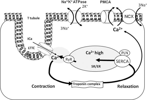
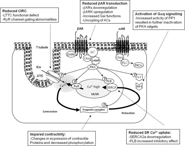
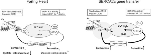
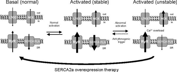
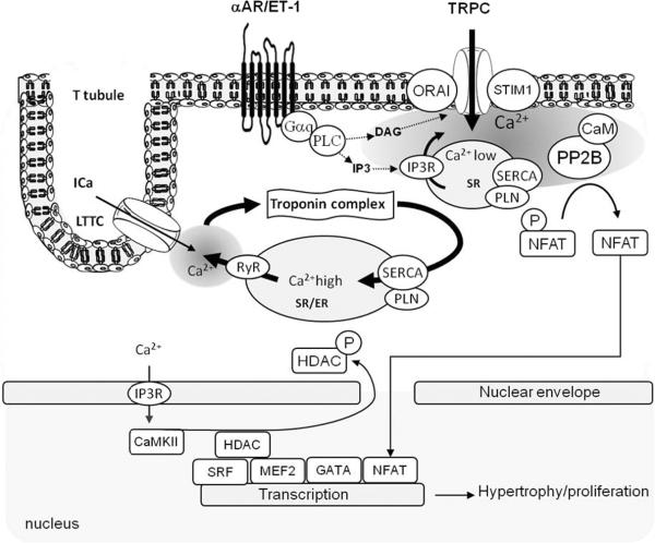
The cardiac isoform of the sarco/endoplasmic reticulum Ca(2+)ATPase (SERCA2a) plays a major role in controlling excitation/contraction coupling. In both experimental and clinical heart failure, SERCA2a expression is significantly reduced which leads to abnormal Ca(2+) handling and deficient contractility. A large number of studies in isolated cardiac myocytes and in small and large animal models of heart failure showed that restoring SERCA2a expression by gene transfer corrects the contractile abnormalities and improves energetics and electrical remodeling. Following a long line of investigation, a clinical trial is underway to restore SERCA2a expression in patients with heart failure using adeno-associated virus type 1. This review addresses the following issues regarding heart failure gene therapy: i) new insights on calcium regulation by SERCA2a; ii) SERCA2a as a gene therapy target in animal models of heart failure; iii) advances in the development of viral vectors and gene delivery; and iv) clinical trials on heart failure using SERCA2a. This review focuses on the new advances in SERCA2a- targeted gene therapy made in the last three years. In conclusion, SERCA2a is an important therapeutic target in various cardiovascular disorders. Ongoing clinical gene therapy trials will provide answers on its safety and applicability.


Figures

Figure 1
Figure 1. Excitation-contraction coupling in cardiomyocytes
A small amount of Ca2+ uptake via LTCC causes a large amount of Ca2+ release from the SR via RyR – Ca2+-induced Ca2+ release. Rise of intracellular Ca2+ causes contraction of myofilaments. Ca2+ uptake into the SR by SERCA or extrusion of Ca2+ trough NCX channels results in relaxation of myofilaments. Abbreveations: Na-K ATPase: plasma membrane sodium-potassium ATPase; Ca2+: calcium ion; Ica: L-type calcium current; LTCC: L-type calcium channel; K+: potassium ion; PLN: Phospholamban; PMCA: Plasma Membrane Calcium ATPase; RyR: Ryanodine Receptor calcium channel; Na+: sodium ion; NCX: - sodium-calcium exchanger; SR/ER: Sarco/Endoplasmic Reticulum; SERCA: Sarco/Endoplasmic Reticulum Ca2+ ATPase 2a.
Figure 2
Figure 2. The regulation of calcium cycling via adrenergic pathway and its alterations in failing heart
Schematic showing the positive control of cardiac function through classic β-adrenergic pathway and a negative control of cardiac function through the α-adrenergic pathway. In response to stress, binding of agonist to the βARs results in inotrope (increased contractility), chronotrope (increased heart rate) and lusiotrope (enhanced relaxation) effects. These effects are mediated by PKA phosphorylation of several targets, resulting in enhanced Ca2+ cycling. Negative control of heart function through αARs is mediated by PP1 dephosphorylation of several PKA targets. Abbreveations: αAR: α-adrenergic receptor; AC: Adenylyl cyclase; βAR: β-adrenergic receptor; βARK: β-adrenergic receptor kinase; CIRC: Calcium induced calcium release; Gαq: Gq protein α-subunit; Gαs: Stimulatory G-protein α-subunit; I-1: Inhibitor 1; Ica: L-type calcium current; LTCC: L-type calcium channel; P: Phosphorylation; PKA: Protein kinase A; PKC: Protein kinase C; PLN: Phospholamban; PLC: Phospholipase C; PP1: Serine/threonine phosphatase 1; RyR: Ryanodine receptor calcium channel; SERCA: Sarco/endoplasmic reticulum Ca2+ ATPase.
Figure 3
Figure 3. Effect of SERCA2a overexpression on contractile function in diseased heart
The overexpession of SERCA2a in failing hearts has been shown to result in the recovery of contractility. SERCA2a overexpression improves calcium cycling and myocyte shortening. Abbreveations: CIRC: Calcium induced calcium release; L-type calcium current; LTCC: L-type calcium channel; PLN: Phospholamban; RyR: Ryanodine receptor calcium channel; SERCA: Sarco/endoplasmic reticulum Ca2+ ATPase.
Figure 4
Figure 4. Model showing the effect of SERCA2a overexpression on arrhythmias
The development of an unstable equilibrium is the mechanistic basis of increased arrhythmogenic risk. At the basal level (left) Ca2+ homeostasis is maintained by the balanced activities of LTCC and NCX, and between RyR2 and SERCA2a. Normal cardiac activation (middle) proportionally increases LTCC, NCX, RyR2 and SERCA function to maintain synchronized Ca2+ fluxes resulting in a stable state. Acute disruption of a single component relative to others (e.g. RyR) results in the ablation of synchronized Ca2+ fluxes and directly increases the arrhythmogenic propensity of the system (right). This state, illustrated using the experimentally observed findings of increased RyR2 leak and NCX activity and diminished SERCA activity, is inherently more prone to destabilization. SERCA2a gene transfer removes Ca2+ overload, stabilizes RyR2 and prevents arrhythmias. Abbreveations: Ca2+: calcium ion; LTCC: L-type calcium channel; PLN: Phospholamban; RyR: Ryanodine Receptor calcium channel; NCX: - sodium-calcium exchanger; SR: Sarcoplasmic Reticulum; SERCA: Sarco/Endoplasmic Reticulum Ca2+ ATPase 2a.
Figure 5
Figure 5. Model showing Ca2+ dependent hyperthrophy/proliferation transcription pathways
Excitation/contraction calcium cycling is not involved in the control of hyperthrophic/proliferation pathways. Cardiac myocytes also contain lipid rafts rich in caveolin in the sarcolemma and T-tubules that are thought to generate local microdomains of Ca2+. Activation of phosphoinositol-3-phosphate-coupled membrane receptors results in calcium release from the SR via IP3R. Depletion of SR calcium close to the sarcolemma induces the formation of Store Operated Calcium complex resulted in calcium influx in local submembrane area. This calcium activates calcineurin (PP2B), which dephosphorylates NFAT, inducing its nuclear translocation and transcriptional activation.

Similar articles

Cited by

References

    1. Rosamond W, Flegal K, Furie K, Go A, Greenlund K, Haase N, et al. Heart disease and stroke statistics--2008 update: a report from the American Heart Association Statistics Committee and Stroke Statistics Subcommittee. Circulation. 2008 Jan 29;117(4):e25–146. - PubMed
    1. Jessup M, Brozena S. Heart failure. N Engl J Med. 2003 May 15;348(20):2007–18. - PubMed
    1. Lipskaia L, Ly H, Kawase Y, Hajjar R, Lompre AM. Treatment of heart failure by calcium cycling gene therapy. Future Cardiology. 2007;3(4):413–23. - PubMed
    1. Del Monte F, Hajjar RJ. Intracellular devastation in heart failure. Heart Fail Rev. 2008 Jun;13(2):151–62. - PubMed
    1. Kawase Y, Hajjar RJ. The cardiac sarcoplasmic/endoplasmic reticulum calcium ATPase: a potent target for cardiovascular diseases. Nat Clin Pract Cardiovasc Med. 2008 Sep;5(9):554–65. - PubMed
    2. **Review on calcum cycling gene therapy

Publication types

Substances