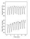
Changes in oxygen partial pressure of brain tissue in an animal model of obstructive apnea
- PMID: 20078851
- PMCID: PMC2817656
- DOI: 10.1186/1465-9921-11-3
Changes in oxygen partial pressure of brain tissue in an animal model of obstructive apnea
Abstract
Background: Cognitive impairment is one of the main consequences of obstructive sleep apnea (OSA) and is usually attributed in part to the oxidative stress caused by intermittent hypoxia in cerebral tissues. The presence of oxygen-reactive species in the brain tissue should be produced by the deoxygenation-reoxygenation cycles which occur at tissue level during recurrent apneic events. However, how changes in arterial blood oxygen saturation (SpO2) during repetitive apneas translate into oxygen partial pressure (PtO2) in brain tissue has not been studied. The objective of this study was to assess whether brain tissue is partially protected from intermittently occurring interruption of O2 supply during recurrent swings in arterial SpO2 in an animal model of OSA.
Methods: Twenty-four male Sprague-Dawley rats (300-350 g) were used. Sixteen rats were anesthetized and non-invasively subjected to recurrent obstructive apneas: 60 apneas/h, 15 s each, for 1 h. A control group of 8 rats was instrumented but not subjected to obstructive apneas. PtO2 in the cerebral cortex was measured using a fast-response oxygen microelectrode. SpO2 was measured by pulse oximetry. The time dependence of arterial SpO2 and brain tissue PtO2 was carried out by Friedman repeated measures ANOVA.
Results: Arterial SpO2 showed a stable periodic pattern (no significant changes in maximum [95.5 +/- 0.5%; m +/- SE] and minimum values [83.9 +/- 1.3%]). By contrast, brain tissue PtO2 exhibited a different pattern from that of arterial SpO2. The minimum cerebral cortex PtO2 computed during the first apnea (29.6 +/- 2.4 mmHg) was significantly lower than baseline PtO2 (39.7 +/- 2.9 mmHg; p = 0.011). In contrast to SpO2, the minimum and maximum values of PtO2 gradually increased (p < 0.001) over the course of the 60 min studied. After 60 min, the maximum (51.9 +/- 3.9 mmHg) and minimum (43.7 +/- 3.8 mmHg) values of PtO2 were significantly greater relative to baseline and the first apnea dip, respectively.
Conclusions: These data suggest that the cerebral cortex is partially protected from intermittently occurring interruption of O2 supply induced by obstructive apneas mimicking OSA.
Figures


Similar articles
-
Tissue oxygenation in brain, muscle, and fat in a rat model of sleep apnea: differential effect of obstructive apneas and intermittent hypoxia.Sleep. 2011 Aug 1;34(8):1127-33. doi: 10.5665/SLEEP.1176. Sleep. 2011. PMID: 21804675 Free PMC article.
-
Brain tissue hypoxia and oxidative stress induced by obstructive apneas is different in young and aged rats.Sleep. 2014 Jul 1;37(7):1249-56. doi: 10.5665/sleep.3848. Sleep. 2014. PMID: 25061253 Free PMC article.
-
Placental oxygen transfer reduces hypoxia-reoxygenation swings in fetal blood in a sheep model of gestational sleep apnea.J Appl Physiol (1985). 2019 Sep 1;127(3):745-752. doi: 10.1152/japplphysiol.00303.2019. Epub 2019 Aug 1. J Appl Physiol (1985). 2019. PMID: 31369330
-
Capillary oxygen saturation and tissue oxygen pressure in the rat cortex at different stages of hypoxic hypoxia.Neurol Res. 2000 Oct;22(7):721-6. doi: 10.1080/01616412.2000.11740746. Neurol Res. 2000. PMID: 11091979
-
Experimental assessment of oxygen homeostasis during acute hemodilution: the integrated role of hemoglobin concentration and blood pressure.Intensive Care Med Exp. 2017 Dec;5(1):12. doi: 10.1186/s40635-017-0125-6. Epub 2017 Mar 1. Intensive Care Med Exp. 2017. PMID: 28251580 Free PMC article.
Cited by
-
Tissue oxygenation in brain, muscle, and fat in a rat model of sleep apnea: differential effect of obstructive apneas and intermittent hypoxia.Sleep. 2011 Aug 1;34(8):1127-33. doi: 10.5665/SLEEP.1176. Sleep. 2011. PMID: 21804675 Free PMC article.
-
Male fertility is reduced by chronic intermittent hypoxia mimicking sleep apnea in mice.Sleep. 2014 Nov 1;37(11):1757-65. doi: 10.5665/sleep.4166. Sleep. 2014. PMID: 25364071 Free PMC article.
-
Age protects from harmful effects produced by chronic intermittent hypoxia.J Physiol. 2016 Mar 15;594(6):1773-90. doi: 10.1113/JP270878. Epub 2016 Feb 9. J Physiol. 2016. PMID: 26752660 Free PMC article.
-
The polymorphic and contradictory aspects of intermittent hypoxia.Am J Physiol Lung Cell Mol Physiol. 2014 Jul 15;307(2):L129-40. doi: 10.1152/ajplung.00089.2014. Epub 2014 May 16. Am J Physiol Lung Cell Mol Physiol. 2014. PMID: 24838748 Free PMC article. Review.
-
Obstructive sleep apnea: impact of hypoxemia on memory.Sleep Breath. 2013 May;17(2):811-7. doi: 10.1007/s11325-012-0769-0. Epub 2012 Oct 13. Sleep Breath. 2013. PMID: 23065547 Free PMC article.
References
-
- Caples SM, Garcia-Touchard A, Somers VK. Sleep-disordered breathing and cardiovascular risk. Sleep. 2007;30:291–303. - PubMed

