. 2010;12(1):R8.
doi: 10.1186/bcr2473. Epub 2010 Jan 15.

(-)-Epigallocatechin gallate sensitizes breast cancer cells to paclitaxel in a murine model of breast carcinoma

Affiliations

(-)-Epigallocatechin gallate sensitizes breast cancer cells to paclitaxel in a murine model of breast carcinoma

Ting Luo et al. Breast Cancer Res. 2010.

Abstract

Introduction: Paclitaxel (Taxol) is a microtubule-targeted agent that is widely used for cancer treatment. However, resistance to paclitaxel is frequently encountered in the clinic. There is increasing interest in identifying compounds that may increase the sensitivity to conventional chemotherapeutic agents. In this study, we investigated whether green tea polyphenol (-)-epigallocatechin gallate (EGCG) could sensitize breast carcinoma to paclitaxel in vivo.

Methods: Breast cancer cells were treated with or without EGCG and paclitaxel followed by detection of cell survival and apoptosis. c-Jun NH2-terminal kinase (JNK) phosphorylation and glucose-regulated protein 78 (GRP78) expression were detected by Western blotting. For in vivo study, 4T1 breast cancer cells were inoculated into Balb/c mice to establish a transplantation model. The tumor-bearing mice were treated with or without EGCG (30 mg/kg, i.p.) and paclitaxel (10 mg/kg, i.p.). Tumor growth was monitored. Apoptosis in tumor tissues was detected. Cell lysates from tumors were subjected to Western blot analysis of GRP78 expression and JNK phosphorylation.

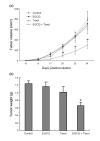
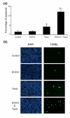
Results: EGCG synergistically sensitized breast cancer cells to paclitaxel in vitro and in vivo. EGCG in combination with paclitaxel significantly induced 4T1 cells apoptosis compared with each single treatment. When tumor-bearing mice were treated with paclitaxel in combination with EGCG, tumor growth was significantly inhibited, whereas the single-agent activity for paclitaxel or EGCG was poor. EGCG overcame paclitaxel-induced GRP78 expression and potentiated paclitaxel-induced JNK phosphorylation in 4T1 cells both in vitro and in vivo.

Conclusions: EGCG may be used as a sensitizer to enhance the cytotoxicity of paclitaxel.


Figures

Figure 1
Figure 1
EGCG sensitizes breast cancer cells to taxol. (a) Sensitivity of 4T1, MDA-MB-231 and MCF-7 cells to taxol. Cells were treated with increasing doses of taxol (0.1 to 2 μM) for 24 h. Cell viability was assessed by WST1 assay. Points = mean of four replicates; bars = SE. Asterisk = P < 0.01 versus untreated controls. (b) 4T1 cells were seeded in a 96-well plate at 5,000 cells per well. The next day, the cells were treated with 20 μM EGCG, 1 μM taxol, or a combination for 48 h. Cell viability was assessed by WST1 assay. Points = mean of four replicates; bars = SE. Asterisk = P < 0.01 versus untreated control. Triangle = P < 0.001 versus single agent. (c) MDA-MB-231 cells were seeded in a 96-well plate at 5,000 cells per well. The next day, the cells were treated with 20 μM EGCG, 1 μM taxol, or combination for 48 h. Cell viability was assessed by WST1 assay. Points = mean of four replicates; bars = SE. Asterisk = P < 0.001 versus untreated control. Triangle = P < 0.001 versus single agent.
Figure 2
Figure 2
EGCG potentiates taxol-induced apoptosis in 4T1 cells. (a) 4T1 cells were treated with 20 μM EGCG, 1 μM taxol, 10 μM SP600125 or combination for 24 h, and apoptosis was assessed by Hoechst 33342 staining. The apoptotic cells with strong fluorescence or fragmented nuclei were observed under fluorescent microscopy. (b) Quantification of apoptotic cells was performed by taking the images in random fields and counting cells with strong fluoresence or fragmented nuclei. The relative apoptosis rate was plotted. Columns = mean percentage of apoptotic cells; bars = SE. Asterisk = P < 0.001 versus untreated control or taxol in combination with 10 μM SP600125. Black triangle = P < 0.001 versus EGCG, taxol, or taxol in combination with EGCG and SP600125. (c) 4T1 cells were treated with 20 μM EGCG, 1 μM taxol, EGCG in combination with taxol, taxol in combination with 10 μM SP600125, or taxol in combination with EGCG and SP600125 for 24 h. Total proteins were harvested with lysis buffer containing phosphatase inhibitor, and subjected to Western blot analysis of JNK phosphorylation. The ratio of p-JNK/actin as determined by densitometric analysis was given. (d) 4T1 cells were treated with 20 μM EGCG, 1 μM taxol, or EGCG in combination with taxol for 24 h. Total proteins were harvested and subjected to Western blot analysis of GRP78 expression. The ratio of GRP78/actin as determined by densitometric analysis was given. Representative of two experiments was shown.
Figure 3
Figure 3
EGCG sensitizes 4T1 tumors to taxol in Balb/c mice. (a) 4T1 cells were injected into Balb/c mice as described in Materials and Methods. When tumors became palpable, the mice were randomly assigned into different groups (n = 8 for control; n = 8 for EGCG group; n = 8 for taxol group; n = 10 for combination treatment), and treated with EGCG (30 mg/kg), taxol (10 mg/kg), or both. Tumor growth was monitored for 24 days. Asterisk = P < 0.05, significantly different from control, EGCG, or taxol group (two-way ANOVA, post-hoc test). Data are mean ± SE. (b) Tumors were dissected 24 days after inoculation of tumor cells into the mice. The tumor weight was measured. The combined data of tumor weight from two experiments was plotted (n = 15 for control; n = 15 for EGCG group; n = 15 for taxol group; n = 16 for combination treatment). Columns = mean of tumor weight; bars = SE. Asterisk = P < 0.05, significantly different from control or single agent.
Figure 4
Figure 4
EGCG potentiates taxol-induced apoptosis in tumors. (a) Percent apoptosis relative to the saline-treated control tumors. Columns = mean of apoptosis rate in tumors; bars = SE. Asterisk = P < 0.01, significantly different from control. Triangle = P < 0.001, significantly different from taxol alone. (b) Representative TUNEL assays for tumors were shown.
Figure 5
Figure 5
EGCG overcomes taxol-induced GRP78 expression in tumors. Cell lysates from dissected tumor tissues were subjected to Western blot analysis of GRP78 expression. (a) The levels of GRP78 protein in tumors treated with vehicle, EGCG, and taxol were shown. (b) Quantitative analysis of GRP78 expression after normalization to actin by densitometric analysis. The ratio of GRP78/actin represented by grey column was set as 1 arbitary unit. Columns = mean of relative levels of GRP78 protein; bars = SE. Asterisk = P < 0.05 versus control. Black triangle = P < 0.05 versus taxol.
Figure 6
Figure 6
EGCG potentiates taxol-induced JNK phosphorylation in tumors. Cell lysates from dissected tumors were subjected to Western blot analysis of JNK phosphorylation. The levels of phosphorylated JNK in tumors treated with or without EGCG and taxol were shown.

Similar articles

Cited by

References

    1. Rowinsky EK, Donehower RC. Paclitaxel (taxol) N Engl J Med. 1995;332:1004–1014. doi: 10.1056/NEJM199504133321507. - DOI - PubMed
    1. Arnal I, Wade RH. How does taxol stabilize microtubules? Curr Biol. 1995;5:900–908. doi: 10.1016/S0960-9822(95)00180-1. - DOI - PubMed
    1. McGuire WP, Rowinsky EK, Rosenshein NB, Grumbine FC, Ettinger DS, Armstrong DK, Donehower RC. Taxol: a unique antineoplastic agent with significant activity in advanced ovarian epithelial neoplasms. Ann Intern Med. 1989;111:273–279. - PubMed
    1. Schiff PB, Fant J, Horwitz SB. Promotion of microtubule assembly in vitro by taxol. Nature. 1979;277:665–667. doi: 10.1038/277665a0. - DOI - PubMed
    1. Horwitz SB. Mechanism of action of taxol. Trends Pharmacol Sci. 1992;13:134–136. doi: 10.1016/0165-6147(92)90048-B. - DOI - PubMed

Publication types