. 2010 Jan 15:11:3.
doi: 10.1186/1471-2121-11-3.

Modulatory effects of cAMP and PKC activation on gap junctional intercellular communication among thymic epithelial cells

Affiliations

Modulatory effects of cAMP and PKC activation on gap junctional intercellular communication among thymic epithelial cells

Oscar K Nihei et al. BMC Cell Biol. .

Abstract

Background: We investigated the effects of the signaling molecules, cyclic AMP (cAMP) and protein-kinase C (PKC), on gap junctional intercellular communication (GJIC) between thymic epithelial cells (TEC).

Results: Treatment with 8-Br-cAMP, a cAMP analog; or forskolin, which stimulates cAMP production, resulted in an increase in dye transfer between adjacent TEC, inducing a three-fold enhancement in the mean fluorescence of coupled cells, ascertained by flow cytometry after calcein transfer. These treatments also increased Cx43 mRNA expression, and stimulated Cx43 protein accumulation in regions of intercellular contacts. VIP, adenosine, and epinephrine which may also signal through cyclic nucleotides were tested. The first two molecules did not mimic the effects of 8-Br-cAMP, however epinephrine was able to increase GJIC suggesting that this molecule functions as an endogenous inter-TEC GJIC modulators. Stimulation of PKC by phorbol-myristate-acetate inhibited inter-TEC GJIC. Importantly, both the enhancing and the decreasing effects, respectively induced by cAMP and PKC, were observed in both mouse and human TEC preparations. Lastly, experiments using mouse thymocyte/TEC heterocellular co-cultures suggested that the presence of thymocytes does not affect the degree of inter-TEC GJIC.

Conclusions: Overall, our data indicate that cAMP and PKC intracellular pathways are involved in the homeostatic control of the gap junction-mediated communication in the thymic epithelium, exerting respectively a positive and negative role upon cell coupling. This control is phylogenetically conserved in the thymus, since it was seen in both mouse and human TEC preparations. Lastly, our work provides new clues for a better understanding of how the thymic epithelial network can work as a physiological syncytium.


Figures
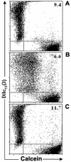
Figure 1
Figure 1
Flow cytometric analysis of the mouse thymic epithelial cell line, showing inter-TEC gap junction intercellular communication. Calcein+Dilc18(3)- and calcein-DiIc18(3)+ IT-76M1 cells were co-cultured for 6 hr at 37°C. These cells were then dissociated and analyzed by flow cytometry to quantify the double positive [calcein+Dilc18(3)+] cells. Some calcein+Dilc18(3)- and calcein-DiIc18(3)+ cells were separately cultured and used to adjust the cytometry settings. These cells also were used to establish the control population (A). Data are presented in the form of dot plots (A, B, C), which depict two-dimensionally the labeling pattern of each cell population considering the fluorescence intensity (log scale) of calcein and DiIc18(3). In B, the 6 hr co-cultured cells are shown, where the presence of double positive cells is apparent, indicating the dye coupling. In C, cells co-cultured for 6 hours in the presence of 18-β-glycyrrhetinic acid (GRA; 100 μM) exhibited a complete inhibition of inter-TEC GJIC. These data are representative of at least 4 experiments.
Figure 2
Figure 2
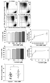
Increase in cAMP enhances inter-TEC GJIC. Panels A and B depict untreated co-cultures of the mouse TEC line at zero and 6 hours time points, respectively. The percentage of double positive cells and the calcein geometric mean fluorescence intensity (MFI) of these populations are depicted at the upper right corner of each panel (%, above; MFI, below). Panels C and D show the inter-TEC coupling, following 6 hours of treatment with 1 mM 8-Br-cAMP or 10 μM forskolin. Both treatments enhanced the calcein mean fluorescence intensity of coupled cells (double positive cells). These data are representative of at least 4 separate experiments. Such enhancements can also be seen in Panels E to H, depicting TEC co-cultures treated for 6 hours with increasing concentrations of either 8-Br-cAMP (E-F) or forskolin (G-H). While percentages of coupled TEC was not significantly modified (E, G), the geometric mean fluorescence of calcein quantified from the double positive cells tripled after both treatments (F, H). The results are representative of 3 independent experiments (mean SD). Panel I shows that 8-Br-cAMP was also capable of enhancing inter-TEC GJIC, in primary cultures of human TNC-derived epithelial cells. Numbers of coupled cells were count in blind. * p < 0.05.
Figure 3
Figure 3
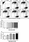
Vasoactive intestinal peptide (VIP), adenosine and epinephrine effects on basal levels of inter-TEC GJIC. Co-cultures of the mouse TEC line were either treated or not (Ct- 6 hrs) with increasing concentrations of VIP (1 - 1000 nM, panels C to F), and the degree of cell coupling, ascertained by cytofluorometry, did not change as compared to the 6 hours untreated control (B), in relation to both percentages of coupled cells and calcein mean fluorescence. Values are shown at the upper right corner of each panel, representing the percentage of double positive cells (above) and the calcein geometric mean fluorescence intensity (below) of these populations. Panel A depicts the flow cytometry profiles of TEC that were not co-cultured. Panel G shows that adenosine does not alter inter-TEC GJIC as well, as revealed by the percentages of coupled cells seen after treatment of various doses of the nucleotide. Panel H shows that epinephrine (1 nM to 10 μM) increased the percentage of dye coupling between TEC in a dose-dependent fashion. Data are shown as mean ± standard deviation, being representative of 2 independent experiments performed in triplicate.
Figure 4
Figure 4
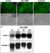
Increase in cAMP enhances connexin gene and protein expression. Cultures of the mouse TEC line were treated with either 8-Br-cAMP (1 mM) or forskolin (10 μM) for 6 hrs at 37°C. Cells were then fixed, permeabilized and labeled with the anti-Cx43 polyclonal antibody, ultimately revealed with the Alexa 488-conjugated secondary antibody. The fluorescence microscopy images (A, C, E) and the corresponding phase contrast images are depicted (B, D, F). The mouse TEC treated with 8-Br-cAMP (C, D) and forskolin (E, F) presented an increased punctate labeling of Cx43, mainly at cell-to-cell contact regions, when compared with the untreated controls (A, B). Inserts in A and B show the fluorescence microscopy image and respective phase contrast image of TEC subjected to isotype control primary antibody and Alexa-488-coupled secondary antibody (Magnification, ×400). Panel G show by northern blot analysis that 8-Br-cAMP also enhances Cx43 gene transcription. Mouse TEC were treated or not (Ct) with 8-Br-cAMP (1 mM) and cultured for 1, 6 or 24 hrs at 37°C. The total RNA was then extracted, and 10 - 20 μg of total RNA was loaded in 1.2% agarose gel and plotted onto nylon membrane. For detection of Cx43 mRNA, the membrane was hybridized with a 32P-labeled cDNA probe for Cx43. Alternatively, the membrane was also hybridized with a 32P-labeled cDNA for GAPDH. The amount of Cx43 mRNA in IT-76M1 cells was increased after 1, 6, and 24 hours of treatment with 8-Br-cAMP.
Figure 5
Figure 5
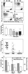
Phorbol myristate acetate inhibits GJIC in mouse and human TEC. Panels A to D are flow cytometry profiles showing mouse TEC co-cultures that were either treated (C) or not (B) with PMA (10 ng/ml) and maintained for 6 hrs at 37°C. Panel A represents the TEC population, which was separately cultured (Ct - 0 hours). The percentage of double positive cells is depicted at the upper right corner of each panel. The histograms with the calcein fluorescence profile of each population are depicted in panel D: calcein-Dilc18(3)+ cells, not submitted to co-culture (gray filled profile); control co-cultured TEC (black line); co-cultured TEC treated with PMA (gray line). Data are representative of at least 4 experiments. Panel E shows a dose-response curve of the effect of PMA treatment upon inter-TEC GJIC. A significant dose-dependent inhibition of cell coupling is seen, with a plateau being reached in 100 ng/ml. (* p < 0.05). Panel F shows that PMA also down-regulates GJIC in primary cultures of TNC-derived human TEC. Panel G shows that simultaneous treatment with PMA and ionomycin also significantly inhibited dye coupling among TEC (* p < 0.05). Co-cultures of the mouse TEC line were treated simultaneously with PMA (10 or 100 ng/ml) and ionomycin (1 μg/ml) for 6 hrs at 37°C, analyzed by flow cytometry. The percentage of coupled cells and the calcein geometric mean fluorescence obtained from double positive cells (Mean ± SD) are shown. The data are representative of two independent experiments performed in triplicate.
Figure 6
Figure 6
The presence of thymocytes do not change inter-TEC GJIC. Mouse TEC co-cultures (IT-76M1 cells) were maintained at 37°C for 2 hrs to establish an adherent confluent monolayer, and then simultaneously cultured with murine thymocytes at 1:5 or 1:10 (TEC:thymocytes) proportion for additional 5 hrs. After this incubation the thymocytes were discarded and epithelial cells were dissociated and analyzed by flow cytometry. The normalized dye coupling degree (gray columns) and the calcein mean fluorescence (white columns) of the double positive cells, clearly show the presence of thymocytes did not significantly modify the levels of dye coupling and dye transfer efficiency among mouse TEC. Data are expressed as mean SD, being derived from obtained from three independent experiments.

Similar articles

Cited by

References

    1. Caveney S. The role of gap junctions in development. Annu Rev Physiol. 1985;47:319–335. doi: 10.1146/annurev.ph.47.030185.001535. - DOI - PubMed
    1. Decrock E, Vinken M, De VE, Krysko DV, D'Herde K, Vanhaecke T, Vandenabeele P, Rogiers V, Leybaert L. Connexin-related signaling in cell death: to live or let die? Cell Death Differ. 2009;16:524–536. doi: 10.1038/cdd.2008.196. - DOI - PubMed
    1. Hertzberg EL, Lawrence TS, Gilula NB. Gap junctional communication. Annu Rev Physiol. 1981;43:479–491. doi: 10.1146/annurev.ph.43.030181.002403. - DOI - PubMed
    1. Kardami E, Dang X, Iacobas DA, Nickel BE, Jeyaraman M, Srisakuldee W, Makazan J, Tanguy S, Spray DC. The role of connexins in controlling cell growth and gene expression. Prog Biophys Mol Biol. 2007;94:245–264. doi: 10.1016/j.pbiomolbio.2007.03.009. - DOI - PubMed
    1. Loewenstein WR. Junctional intercellular communication: the cell-to-cell membrane channel. Physiol Rev. 1981;61:829–913. - PubMed

LinkOut - more resources