. 2010 Jan 15:11:36.
doi: 10.1186/1471-2164-11-36.

The proteolytic system of lactic acid bacteria revisited: a genomic comparison

Affiliations

The proteolytic system of lactic acid bacteria revisited: a genomic comparison

Mengjin Liu et al. BMC Genomics. .

Abstract

Background: Lactic acid bacteria (LAB) are a group of gram-positive, lactic acid producing Firmicutes. They have been extensively used in food fermentations, including the production of various dairy products. The proteolytic system of LAB converts proteins to peptides and then to amino acids, which is essential for bacterial growth and also contributes significantly to flavor compounds as end-products. Recent developments in high-throughput genome sequencing and comparative genomics hybridization arrays provide us with opportunities to explore the diversity of the proteolytic system in various LAB strains.

Results: We performed a genome-wide comparative genomics analysis of proteolytic system components, including cell-wall bound proteinase, peptide transporters and peptidases, in 22 sequenced LAB strains. The peptidase families PepP/PepQ/PepM, PepD and PepI/PepR/PepL are described as examples of our in silico approach to refine the distinction of subfamilies with different enzymatic activities. Comparison of protein 3D structures of proline peptidases PepI/PepR/PepL and esterase A allowed identification of a conserved core structure, which was then used to improve phylogenetic analysis and functional annotation within this protein superfamily.The diversity of proteolytic system components in 39 Lactococcus lactis strains was explored using pangenome comparative genome hybridization analysis. Variations were observed in the proteinase PrtP and its maturation protein PrtM, in one of the Opp transport systems and in several peptidases between strains from different Lactococcus subspecies or from different origin.

Conclusions: The improved functional annotation of the proteolytic system components provides an excellent framework for future experimental validations of predicted enzymatic activities. The genome sequence data can be coupled to other "omics" data e.g. transcriptomics and metabolomics for prediction of proteolytic and flavor-forming potential of LAB strains. Such an integrated approach can be used to tune the strain selection process in food fermentations.


Figures

Figure 1
Figure 1
Distribution of proteinase, peptide transporters and peptidases of the proteolytic system in LAB. The number of identified genes is indicated. MEROPS families are indicated for proteinase and peptidases. Color shading shows absence of a gene (white), a single gene (yellow) or multiple genes (green). The GI codes of the genes can be found in Additional File 2.
Figure 2
Figure 2
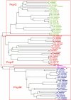
Superfamily tree of PepP/PepQ/PepM members in LAB. Genome abbreviations can be found in "Methods". For each gene, the organism abbreviations are followed by GI codes. Homologs from two non-LAB strains are also included, CBO for Clostridium botulinum F str. Langeland and ECO for E. coli. Experimentally characterized genes are highlighted by the red dots. Green circles represent the speciation events, and red squares represent duplication events.
Figure 3
Figure 3
Superfamily tree of PepD members in LAB. PepD that is experimentally characterized from Bifidobacterium longum NCC2705 (BLO) [52] and pepD genes from L. helveticus CNRZ32 (LHV) analyzed by microarray [32] are indicated by the red dots. Green circles represent the speciation events, while red squares represent duplication events.
Figure 4
Figure 4
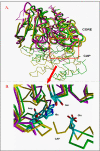
Superposition of 3D structures of proline iminopeptidases 1WM1 (yellow) and 1MTZ (green), and esterase 2UZ0 (purple). The structure of 1AZW is highly similar to 1WM1 and is not shown. A) The 4 conserved structural core segments are shown as thick tubes, and the variable segments as thin sticks connecting C-alpha atoms. The variable large cap regions of the peptidases, which do not superpose, are at the bottom half of the figure. Note that the esterase has a much shorter connecting segment in this cap region. The red frame indicates the position of the active site, which is shown as the zoomed-in view in Panel B. B) The catalytic site is shown with catalytic residues Ser, His and Asp. The active site is enlarged and rotated by about 180 degrees relative to Panel A. A short stretch of the cap region in both peptidases is shown, bearing the Glu residues that interact with the positive charge of the peptide substrate N-terminus. Note that the side chains of the two Glu residues superpose very well, despite coming from different (non-superposable) parts of the cap region.
Figure 5
Figure 5
Superfamily tree of PepI/PepR/PepL and EstA members. Based on MSA of the concatenated sequences of the four structural core regions identified by the protein 3D structure alignment. Orthologous proteins are indicated by the same color. The PepR subgroup from LAB is shadowed in pink, and the PepL subgroup is shadowed in yellow. The bacterial phyla are indicated. Red dots indicate the experimentally characterized genes and red triangles indicate the protein 3D structures used for the analyses. The event of the substitution of catalytic residue aspartate by glutamate in the PepR subgroup is indicated in the tree.

Similar articles

Cited by

References

    1. Christensen JE, Dudley EG, Pederson JA, Steele JL. Peptidases and amino acid catabolism in lactic acid bacteria. Antonie Van Leeuwenhoek. 1999;76:217–246. doi: 10.1023/A:1002001919720. - DOI - PubMed
    1. Kim YK, Yaguchi M, Rose D. Isolation and Amino Acid Composition of Para-Kappa-Casein. J Dairy Sci. 1969;52:316–320.
    1. Stewart AF, Bonsing J, Beattie CW, Shah F, Willis IM, Mackinlay AG. Complete nucleotide sequences of bovine alpha S2- and beta-casein cDNAs: comparisons with related sequences in other species. Mol Biol Evol. 1987;4:231–241. - PubMed
    1. Liu M, Nauta A, Francke C, Siezen RJ. Comparative genomics of enzymes in flavor-forming pathways from amino acids in lactic acid bacteria. Appl Environ Microbiol. 2008;74:4590–4600. doi: 10.1128/AEM.00150-08. - DOI - PMC - PubMed
    1. Kunji ER, Mierau I, Hagting A, Poolman B, Konings WN. The proteolytic systems of lactic acid bacteria. Antonie Van Leeuwenhoek. 1996;70:187–221. doi: 10.1007/BF00395933. - DOI - PubMed

Publication types