. 2010 Jan 15:7:6.
doi: 10.1186/1743-422X-7-6.

Human coronavirus NL63 open reading frame 3 encodes a virion-incorporated N-glycosylated membrane protein

Affiliations

Human coronavirus NL63 open reading frame 3 encodes a virion-incorporated N-glycosylated membrane protein

Marcel A Müller et al. Virol J. .

Abstract

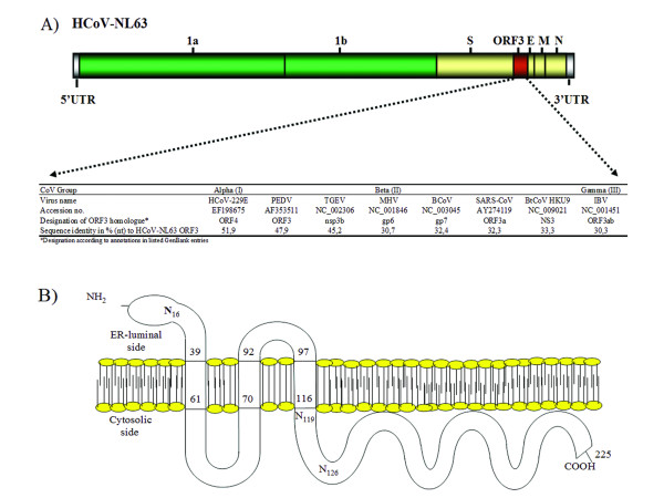
Background: Human pathogenic coronavirus NL63 (hCoV-NL63) is a group 1 (alpha) coronavirus commonly associated with respiratory tract infections. In addition to known non-structural and structural proteins all coronaviruses have one or more accessory proteins whose functions are mostly unknown. Our study focuses on hCoV-NL63 open reading frame 3 (ORF 3) which is a highly conserved accessory protein among coronaviruses.

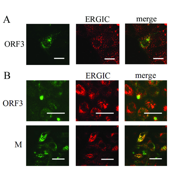
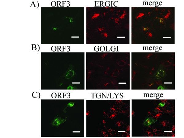
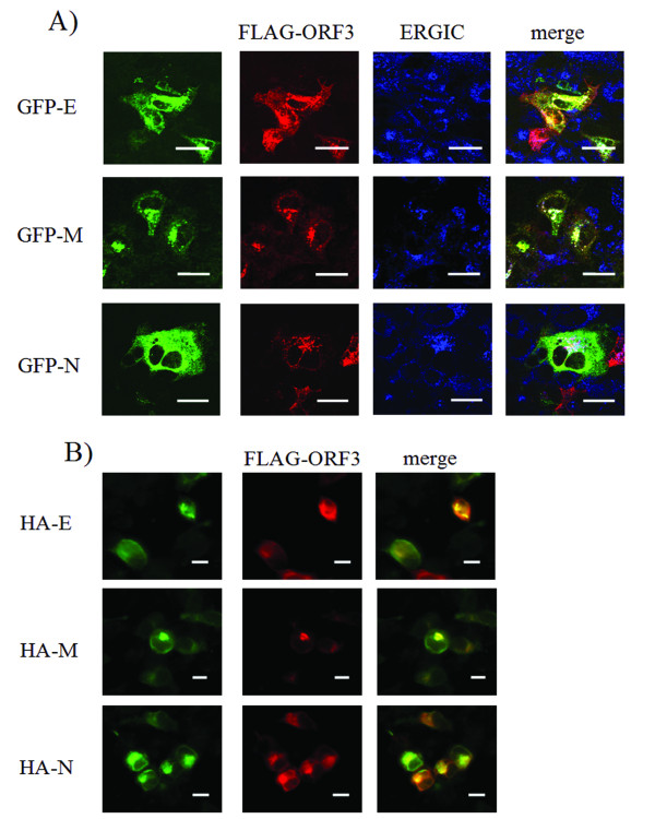
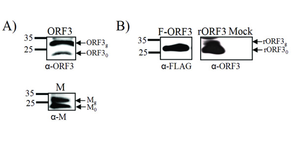
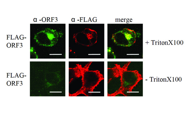
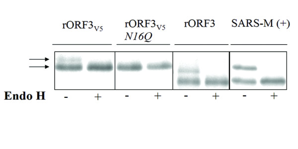
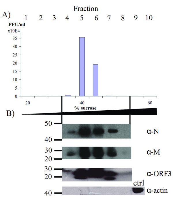
Results: In-silico analysis of the 225 amino acid sequence of hCoV-NL63 ORF 3 predicted a triple membrane-spanning protein. Expression in infected CaCo-2 and LLC-MK2 cells was confirmed by immunofluorescence and Western blot analysis. The protein was detected within the endoplasmatic reticulum/Golgi intermediate compartment (ERGIC) where coronavirus assembly and budding takes place. Subcellular localization studies using recombinant ORF 3 protein transfected in Huh-7 cells revealed occurrence in ERGIC, Golgi- and lysosomal compartments. By fluorescence microscopy of differently tagged envelope (E), membrane (M) and nucleocapsid (N) proteins it was shown that ORF 3 protein colocalizes extensively with E and M within the ERGIC. Using N-terminally FLAG-tagged ORF 3 protein and an antiserum specific to the C-terminus we verified the proposed topology of an extracellular N-terminus and a cytosolic C-terminus. By in-vitro translation analysis and subsequent endoglycosidase H digestion we showed that ORF 3 protein is N-glycosylated at the N-terminus. Analysis of purified viral particles revealed that ORF 3 protein is incorporated into virions and is therefore an additional structural protein.

Conclusions: This study is the first extensive expression analysis of a group 1 hCoV-ORF 3 protein. We give evidence that ORF 3 protein is a structural N-glycosylated and virion-incorporated protein.


Figures

Figure 1
Figure 1
Characteristics of hCoV-NL63 open reading frame 3 and comparison to homologous genes in other coronaviruses. The sequence of ORF 3 (GenBank accession no. AY567487.2) was analyzed using BLAST and MEGA4. (A), localization of ORF 3 within the hCoV-NL63 genome and comparison of nucleotide (nt) identity based on multiple sequence alignments with prototype strains of CoV groups alpha, beta, gamma. Note that IBV ORF 3a and b were fused to one ORF 3ab. (B), Summarized results of in-silico analysis on membrane topology and glycosylation (refer to Materials and Methods section). Predicted N-linked glycosylation sites are indicated by an "N" at the respective localizations with an index number indentifying the amino acid position. No O linked glycosylation sites were predicted.
Figure 2
Figure 2
Subcellular localization of viral proteins in hCoV-NL63 infected CaCo-2 and LLC-MK2 cells by immunofluorescence assay. Confocal laser scanning microscopy on CaCo-2 (A) and LLC-MK2 cells (B) infected with hCoV-NL63. Left panels: staining with anti-ORF 3 and anti-M protein rabbit antisera (only in B) and detection by fluorescein isothiocyanate (FITC)-labelled goat-anti-rabbit antibody (green). Middle panels: detection of co-staining of the same cells with mouse-anti-ERGIC-53 mAB (Axxora) and detection with rhodamine-labelled goat-anti-mouse antibody. Yellow signals in merged pictures (right panels) show colocalization. Bars represent 20 μm.
Figure 3
Figure 3
Subcellular localization study of overexpressed hCoV-NL63 ORF 3 protein in Huh-7 cells. Confocal laser scanning microscopy on cells expressing recombinant ORF 3 protein and co-staining with different antibodies for cellular organelles. Left panels: staining with rabbit-anti-ORF 3 serum and anti-rabbit-Cy2 (Dianova). Middle panels from top to bottom: co-staining of cellular organelles with a mouse-anti-ERGIC53 (A), mouse-anti-Golgi 58 K for the Golgi (B), goat-anti-LAMP-1 for trans-Golgi Network (TGN) and Lysosomes (LYS) together with goat (or donkey)-anti-mouse-Cy3 antibodies (C). Right panels show merged pictures where yellow areas represent colocalization. Partial colocalizations can be observed with all organelle markers indicating that the glycoprotein ORF 3 is processed trans-Golgi. Bars indicate 20 μm.
Figure 4
Figure 4
Subcellular localization of overexpressed hCoV-NL63 proteins in Huh-7 and HEK-293T cells. Confocal laser scanning microscopy on cells co-expressing GFP-E, GFP-M, GFP-N, respectively, together with FLAG-ORF 3. (A), Huh-7 cells. The green panels on the left show GFP fluorescence from overexpressed E, M, and N proteins. Red pictures in the next column show Cy3 fluorescence from anti-FLAG staining of overexpressed FLAG-ORF 3 fusion protein. Blue pictures show Cy5 fluorescence from staining of the ER-Golgi intermediate compartment (ERGIC) (refer to Materials and Methods section for antibodies and staining technique). Yellow areas in the right hand column represent colocalization of the GFP-proteins with FLAG-ORF 3 whereas white regions in merged pictures show colocalization of GFP proteins with FLAG-ORF 3 within the ERGIC. GFP-E and M show excessive colocalization with FLAG-ORF 3 especially within the ERGIC in both cell lines. GFP-N partially colocalizes with FLAG-ORF 3 mainly within the ERGIC. Analysis was performed with the help of a confocal laser scanning microscope (cLSM 510 Meta, Zeiss). Bars represent 20 μm. (B), to exclude altered subcellular localization contributed by the fusion tags on the overexpressed structural proteins, experiments were repeated in HEK-293T cells using FLAG-ORF 3 in combination with HA tagged E, M and N proteins. Bars represent 10 μm.
Figure 5
Figure 5
Comparison of ORF 3 protein in viral infection and overexpression by Western blot. (A), LLC-MK2 cells were inoculated with hCoV-NL63 (MOI 0.01) and analyzed by Western blot after 4 days using antibodies against the ORF 3 protein C-terminus (top) and against M (bottom). The bands named ORF 30 and M0 are corresponding to the predicted molecular weights of both proteins (26 kDa). Larger bands ORF 3 g and Mg were assumed to be the result of posttranslational modification. (B, left panel): HEK-293T cells transfected with N-terminally FLAG-tagged ORF 3 do not show signs of posttranslational modification as observed in (A). (B, right panel): overexpression of ORF 3 protein in the same system without an N-terminal fusion tag reconstitutes the additional band of higher molecular weight observed in infected cells. The "mock" lane represents a control transfected with an empty vector.
Figure 6
Figure 6
Topology of recombinant FLAG-tagged ORF 3 protein. Recombinant N-terminal tagged FLAG-ORF 3 protein was transiently expressed in HEK-293T cells and localization was analyzed by confocal laser scanning microscopy (cLSM 510 Meta, Zeiss). FLAG-ORF 3 protein was stained with rabbit-anti-ORF 3 recognizing the C-terminus and mouse-anti-FLAG for detection of the FLAG-tagged N-terminus (upper panel). Permeabilized cells (+TritonX100) show colocalized signals mainly in perinuclear regions for protein ORF 3 C-terminus and N-terminus whereas without permeabilization (-TritonX100) only FLAG-tagged N-terminus of protein ORF 3 could be detected at the plasma membrane (lower panel). Bars represent 10 μm.
Figure 7
Figure 7
N-glycosylation of hCoV-NL63 ORF 3 protein. HCoV-NL63 ORF 3 protein with and without a C-terminal V5 tag, and with an N16Q exchange in the tagged version was in-vitro translated in presence of 35S-methionine. SARS-CoV M protein without a tag was translated in the same system as a control. Proteins were digested with endoglycosidase (Endo H) as indicated below each lane, subjected to SDS-PAGE, and visualized. Note the removal of the bands of increased molecular weight for the control and ORF 3 proteins, but not for the ORF 3 protein with an amino acid exchange at the hypothetical N-glycosylation site. Note also that extent of size reduction for the SARS-CoV M protein, which is known to have one N-terminal N-glycosylation site, is the same for the NL63 ORF 3 protein.
Figure 8
Figure 8
Identification of NL63-ORF 3 protein as a structural viral protein by sucrose gradient ultracentrifugation. Viral supernatant was purified via subsequently centrifugation on two discontinuous and one continuous sucrose gradients of 20% to 60% (w/v) sucrose. The continuous cushion was divided into ten fractions as indicated in part (A). After centrifugation of each fraction through 20% sucrose cushions, the resulting pellets were analyzed for infectious particles by plaque assays. Resulting virus titers are indicated on the 20 Y-axis in part (A). (B), fractions 4-8 were subjected to Western blot analysis using specific rabbit antibodies against ORF 3, M and N protein (1:3000; 1:250,000 and 1:24,000, respectively). To exclude cellular contaminations in the fractions a Western blot using mouse-anti-actin (1:2,000) was performed. Note the colocalization of the ORF 3 protein in the same gradients as the known structural proteins M and N.

Similar articles

Cited by

References

    1. Hoek L van der, Pyrc K, Jebbink MF, Vermeulen-Oost W, Berkhout RJ, Wolthers KC, Wertheim-van Dillen PM, Kaandorp J, Spaargaren J, Berkhout B. Identification of a new human coronavirus. Nat Med. 2004;7:368–373. doi: 10.1038/nm1024. - DOI - PMC - PubMed
    1. Hofmann H, Pyrc K, Hoek L van der, Geier M, Berkhout B, Pohlmann S. Human coronavirus NL63 employs the severe acute respiratory syndrome coronavirus receptor for cellular entry. Proc Natl Acad Sci USA. 2005;7:7988–7993. doi: 10.1073/pnas.0409465102. - DOI - PMC - PubMed
    1. Han TH, Chung JY, Kim SW, Hwang ES. Human Coronavirus-NL63 infections in Korean children, 2004-2006. J Clin Virol. 2007;7:27–31. doi: 10.1016/j.jcv.2006.10.009. - DOI - PMC - PubMed
    1. Vabret A, Mourez T, Dina J, Hoek L van der, Gouarin S, Petitjean J, Brouard J, Freymuth F. Human coronavirus NL63, France. Emerg Infect Dis. 2005;7:1225–1229. - PMC - PubMed
    1. Pyrc K, Berkhout B, Hoek L van der. The novel human coronaviruses NL63 and HKU1. J Virol. 2007;7:3051–3057. doi: 10.1128/JVI.01466-06. - DOI - PMC - PubMed

Publication types

MeSH terms

LinkOut - more resources