. 2010 Jan 15:11:4.
doi: 10.1186/1471-2199-11-4.

Halogenated imidazole derivatives block RNA polymerase II elongation along mitogen inducible genes

Affiliations

Halogenated imidazole derivatives block RNA polymerase II elongation along mitogen inducible genes

Michal Mikula et al. BMC Mol Biol. .

Abstract

Background: Aberrant activation of protein kinases is one of the essential oncogenic driving forces inherent to the process of tumorigenesis. The protein kinase CK2 plays an important role in diverse biological processes, including cell growth and proliferation as well as in the governing and transduction of prosurvival signals. Increased expression of CK2 is a hallmark of some cancers, hence its antiapoptotic properties may be relevant to cancer onset. Thus, the designing and synthesis of the CK2 inhibitors has become an important pursuit in the search for cancer therapies.

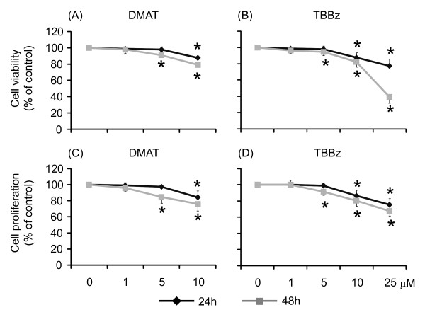
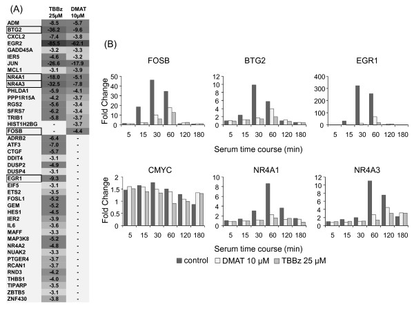
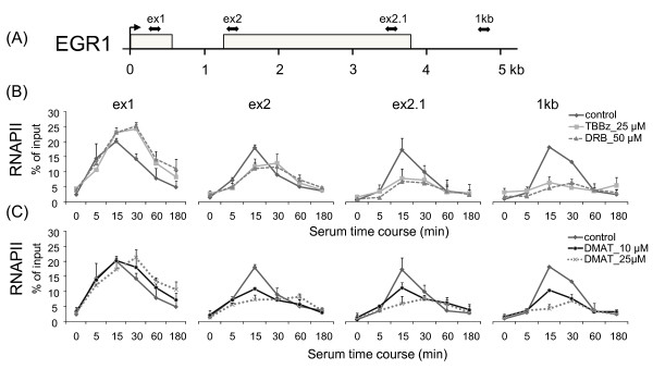
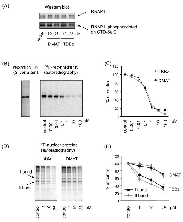
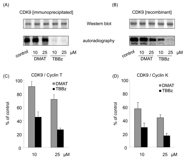
Results: Using a high-throughput microarray approach, we demonstrate that two potent inhibitors of CK2, 4,5,6,7-tetrabromo-benzimidazole (TBBz) and 2-Dimethyloamino-4,5,6,7-tetrabromo-1H-benzimidazole (DMAT), blocked mitogen induced mRNA expression of immediate early genes. Given the impact of these inhibitors on the process of transcription, we investigated their effects on RNA Polymerase II (RNAPII) elongation along the mitogen inducible gene, EGR1 (early growth response 1), using chromatin immunoprecipitation (ChIP) assay. ChIP analysis demonstrated that both drugs arrest RNAPII elongation. Finally, we show that CDK9 kinase activity, essential for the triggering of RNAPII elongation, was blocked by TBBz and to lesser degree by DMAT.

Conclusions: Our approach revealed that small molecules derived from halogenated imidazole compounds may decrease cell proliferation, in part, by inhibiting pathways that regulate transcription elongation.


Figures

Figure 1
Figure 1
The inhibitory effects of DMAT and TBBz on viability and proliferation of HeLa cells. Cells were grown in the presence of 1, 5 and 10 μM of DMAT (A, C) or 1, 5, 10 and 25 μM of TBBz (B, D). Cell viability was monitored by MTT test (A, B), and cell proliferation by 3H thymidine incorporation (C, D) 24 and 48 h later. Four independent experiments were performed, and all assays were repeated in octuplicate. Results are expressed as the percentage of control cell viability or proliferation and represent means ± S.D. *; P < 0.05 compared to the control.
Figure 2
Figure 2
Changes in gene expression induced by TBBz and DMAT. (A) List of 42 genes and fold change in their expression in response to treatment with inhibitors (1 hr) were analyzed by the Affymetrix U133A 2.0 GeneChip microarray. Dark grey indicates a high decrease and light grey lower decrease in expression. Expression of the boxed genes was confirmed by real time PCR and is presented as fold change at zero time (B).
Figure 3
Figure 3
TBBz and DMAT arrest RNAPII elongation along the EGR1 locus after serum treatment. (A) Position of primers relative to transcription start sites (in bp): exon 1 +248; exon 2 +1287; exon 2.1 +3490; 1 kb +4528. (B, C) HeLa cells maintained for 48 h in 0.5% serum were treated with fresh medium supplemented with 15% FBS and -/+ 25 μM TBBz/50 μM DRB/10 or 25 μM DMAT (C) for the indicated times and then used in ChIP assays with antibodies to RNAPII. Purified DNA was used in real-time PCR with pairs of primers spanning the EGR1 locus. The density of RNAPII on the EGR1 gene was quantified by real-time PCR and is presented as a percentage of input. Data represent means ± S.D from 3 independent experiments.
Figure 4
Figure 4
The effects of DMAT and TBBz on phosphorylation of RNAPII and other nuclear proteins. (A) Equal amounts of nuclear extracts prepared from untreated and inhibitor-treated cells (60 min) were separated by SDS-PAGE followed by Western blotting with anti-RNA PII CDT repeat (ab5408, Abcam) or anti-Ser2-phospho-RNAPII CDT (ab5095, Abcam). Similar patterns were obtained in 3 different experiments using different nuclear extracts. (B) Bacterially expressed recombinant hnRNP K protein (rec-hnRNPK, left panel) was phosphorylated by CK2 kinase, purified from rat liver (Sigma; C3460), with increasing concentrations (0; 0.001; 0.01; 0.1; 1.0, 10; 100 μM) of TBBz or DMAT. Assays were stopped by boiling samples in Laemmli loading buffer. K protein was separated by SDS-PAGE, and dried gels were autographed (right panel - representative gel with TBBz treatment). (C) Densitometry of the results with inhibitors are expressed as the percentage of control kinase activity and represent means ± S.D from 3 independent experiments. (D) Nuclear extracts prepared from untreated and inhibitor-treated cells were used for autophosphorylation reactions with or without 1, 10, and 25 μM TBBz or DMAT. Assays were stopped by boiling samples in Laemmli loading buffer. Proteins were separated by SDS-PAGE, and dried gels were autoradiographed (Phosphorimager) (E). Densitometrical analysis of two phosphorylated protein bands [marked on panel (D)] is shown as means ± S.D from results expressed as the percentage of controls.
Figure 5
Figure 5
Differential inhibitory effects of DMAT and TBBz on CDK9 activity in vitro. (A) CDK9/Cyclin T1 proteins pulled down from nuclear extracts (anti-CDK9+anti-cyclin T1, Santa Cruz Biotechnology, D-7 and H-245, respectively) were used in CDK9 autophosphorylation reactions without or with DMAT or TBBz. Autophosphorylated proteins were resolved by SDS-PAGE, transferred to PVDF membrane and immunostained by anti-CDK9 antibody (upper panels) and scanned using a Phosphorimager (lower panels). Similar patterns were obtained in 3 different experiments. (B) Recombinant fusion full-length human CDK9+CyclinK proteins, co-expressed by baculovirus in Sf9 insect cells (Abcam, ab70320) were used in autophosphorylation assays as above. (C) Complexes of CDK9/Cyclin T1 proteins pulled down from nuclear extracts or (D) human CDK9+CyclinK protein were used in kinase assays, with or without DMAT or TBBz. The heptapeptide YSPTSPS was used as a substrate. The reaction mixtures were applied to acidic hydrolysis of γ32P- ATP followed by phosphomolybdate extraction, and 32P-phosphopeptide was determined by liquid scintillation spectrophotometry. Results are expressed as a percentage of kinase inhibition and represent means ± S.D. of 2 separate experiments.

Similar articles

Cited by

References

    1. Hunter T. Signaling--2000 and beyond. Cell. 2000;100(1):113–127. doi: 10.1016/S0092-8674(00)81688-8. - DOI - PubMed
    1. Cohen P. The regulation of protein function by multisite phosphorylation--a 25 year update. Trends in biochemical sciences. 2000;25(12):596–601. doi: 10.1016/S0968-0004(00)01712-6. - DOI - PubMed
    1. Duncan JS, Litchfield DW. Too much of a good thing: the role of protein kinase CK2 in tumorigenesis and prospects for therapeutic inhibition of CK2. Biochimica et biophysica acta. 2008;1784(1):33–47. - PubMed
    1. Kramerov AA, Saghizadeh M, Caballero S, Shaw LC, Li Calzi S, Bretner M, Montenarh M, Pinna LA, Grant MB, Ljubimov AV. Inhibition of protein kinase CK2 suppresses angiogenesis and hematopoietic stem cell recruitment to retinal neovascularization sites. Molecular and cellular biochemistry. 2008;316(1-2):177–186. doi: 10.1007/s11010-008-9831-4. - DOI - PMC - PubMed
    1. Perea SE, Reyes O, Baladron I, Perera Y, Farina H, Gil J, Rodriguez A, Bacardi D, Marcelo JL, Cosme K. CIGB-300, a novel proapoptotic peptide that impairs the CK2 phosphorylation and exhibits anticancer properties both in vitro and in vivo. Molecular and cellular biochemistry. 2008;316(1-2):163–167. doi: 10.1007/s11010-008-9814-5. - DOI - PubMed

Publication types

MeSH terms

LinkOut - more resources