. 2010 May-Jun;1799(5-6):469-79.
doi: 10.1016/j.bbagrm.2010.01.005. Epub 2010 Jan 14.

Estrogen receptor-dependent regulation of CYP2B6 in human breast cancer cells

Affiliations

Estrogen receptor-dependent regulation of CYP2B6 in human breast cancer cells

Raymond Lo et al. Biochim Biophys Acta. 2010 May-Jun.

Abstract

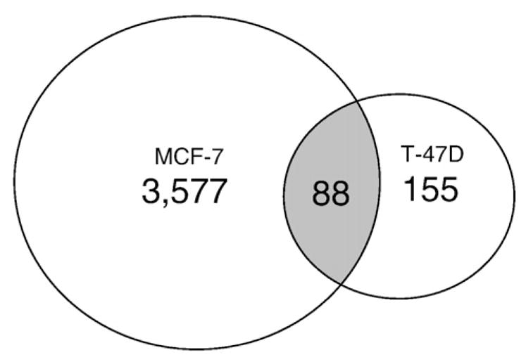
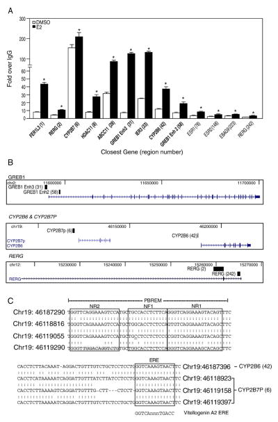
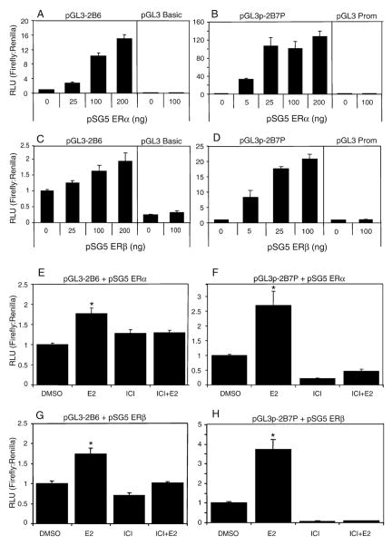
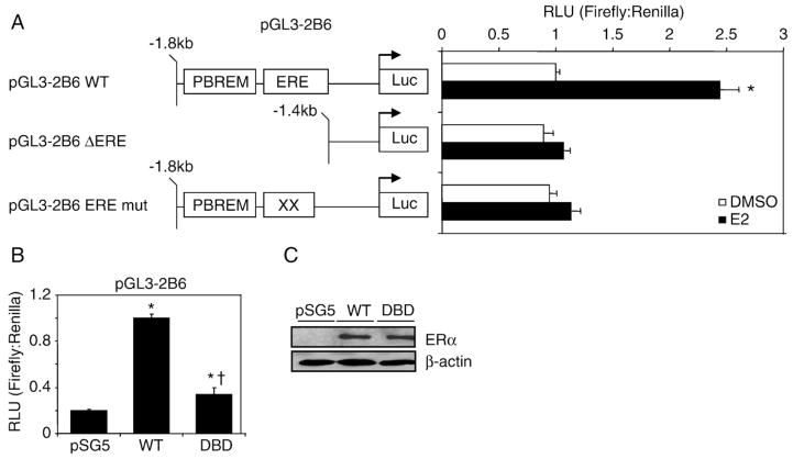
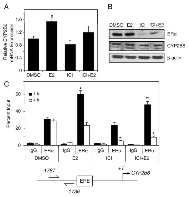
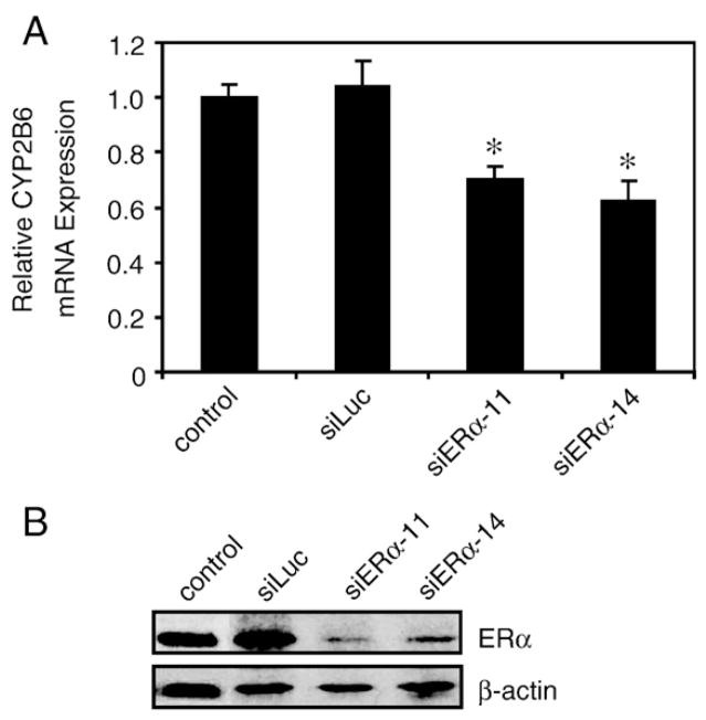
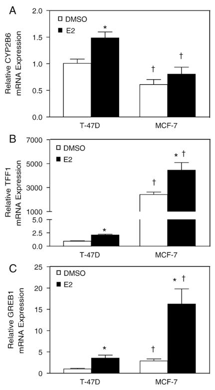
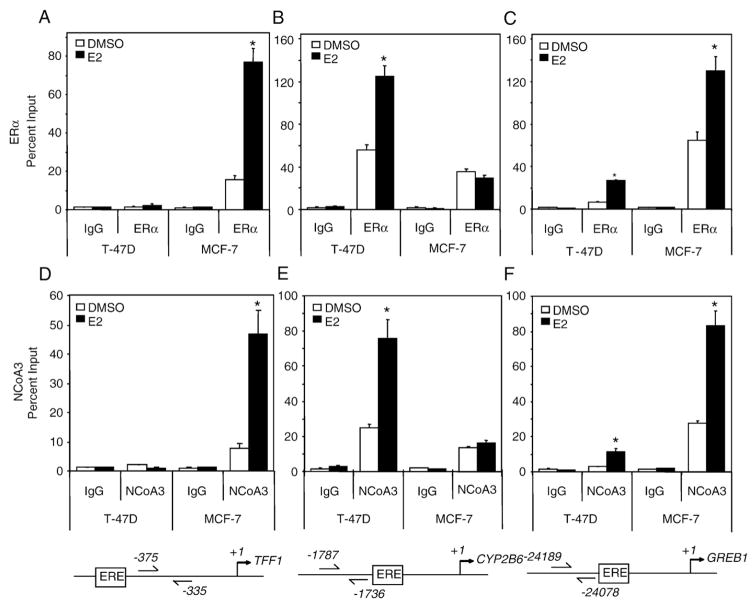
Estrogen receptor alpha (ERalpha) mediates the biological actions of estrogens and also contributes to the development and progression of breast cancer. To gain a more comprehensive understanding of ERalpha-mediated transcription, we used chromatin immunoprecipitation and promoter focused microarrays (ChIP-chip) to identify ERalpha binding sites in T-47D human breast cancer cells. Transcription factor binding site analysis revealed that the estrogen response element (ERE) was significantly over-represented and was found in 50% of the 243 ERalpha-bound regions identified. Interestingly, multiple ERalpha-bound regions were detected in the upstream regulatory sequences of the CYP2B gene cluster. Because ERalpha has been reported to regulate the expression of other cytochrome P450 enzymes and CYP2B6 is highly expressed in ERalpha-positive breast tumors, we focused on characterizing the ERalpha-dependent regulation of CYP2B6. Reporter gene assays revealed that ERalpha and ERbeta increased CYP2B6-regulated gene expression through a functional ERE located at -1669 to -1657 in the upstream regulatory region of CYP2B6. E2 increased ERalpha and nuclear receptor coactivator 3 (NCoA3) recruitment to the 5'-flanking region of CYP2B6, and increased CYP2B6 mRNA levels in T-47D but not in MCF-7 human breast cancer cells. RNAi-mediated knockdown of ERalpha in the T-47D cells resulted in a significant decrease in CYP2B6 mRNA levels. Taken together, our study provides evidence for cell-type specific transcriptional regulation of the CYP2B6 gene by ERs.


Figures

Fig. 1
Fig. 1
ERα-bound regions in T-47D cells. Comparison of the ERα-bound regions identified in the current study (T-47D) with those reported in a genome-wide analysis in MCF-7 cells [27]. As shown in the Venn diagram 88 of the 243 regions identified in our study overlapped with at least 50% sequence identity to those reported previously [27].
Fig. 2
Fig. 2
Confirmation of ERα-bound regions identified in the ChIP-chip study. (A) Conventional ChIP was performed to confirm 13 ERα-bound regions in T-47D plated in DCC-FBS containing medium and treated with E2 for 1 h. These 13 regions were chosen to represent a range of enriched values from the array. ERα recruitment level significantly (p<0.05 Student’s t-test) different from DMSO is indicated by an asterisk for each region. Regions shown in bold font indicate the presence of at least one estrogen response element in the sequence. (B) Selected ERα-bound regions relative to their closest genes are shown using USCS Genome Browser (http://genome.ucsc.edu/). The black blocks represent ERα-bound regions identified from our ChIP-chip study, whereas the arrows indicate the transcriptional direction of the closest annotated genes. (C) The diagram shows the genomic coordinates and the sequences of the putative EREs are boxed in CYP2B7P (6) and CYP2B6 (42) and the phenobarbital-responsive enhancer module (PBREM). Nuclear receptor 1 (NR1) and NR2 represent two direct repeat 4 (DR4) nuclear receptor motifs, which are boxed, while NF1 denotes a putative nuclear factor 1 binding site as previously described [18]. The perfect palindromic ERE from vitellogenin A2 is also shown for comparison. A colon indicates homology among the sequences, where an underline indicates nucleotide differences among the sequences at that position.
Fig. 3
Fig. 3
ER-dependent transcriptional activation of pGL3-2B6 and pGL3p-2B7P in HuH-7 cells. HuH-7 cells were transfected with (A) 200 ng of pGL3-2B6 or (B) pGL3p-2B7P and increasing amounts of the plasmid pSG5 ERα for 24 h prior to luminescence detection. pGL3-2B6 activity is regulated by the natural promoter of CYP2B6, whereas pGL3p-2B7P, by the regulatory region located 69 kb upstream of CYP2B6, which also maps to the 5′ regulatory region of CYP2B7P. Results shown are means±S.E.M. for three independent experiments. Similar results were obtained when increasing amounts of the plasmid pSG5 ERβ were transfected with the two reporter gene plasmids (C and D). In order to evaluate the estrogen responsiveness of CYP2B6, HuH-7 cells were plated in DCC-FBS containing medium for 24 h. Cells were co-transfected with (E) 200 ng of pGL3-2B6 and 50 ng of pSG5 ERα or (F) 200 ng of pGL3p-2B7P and 5 ng of pSG5 ERα for 24 h. The cells were treated with either 0.1% DMSO, 10 nM E2, 100 nM ICI 182,780 or 10 nM E2+100 nM ICI 182,780 for an additional 24 h prior to luminescence detection. Similar results were obtained when (G) 200 ng of pGL3-2B6 and 100 ng of pSG5 ERβ or (H) 200 ng of pGL3p-2B7P and 25 ng of pSG5 ERβ were transfected into the HuH-7 cells. Luciferase activity significantly (p<0.05 one-way ANOVA) different from DMSO is indicated by an asterisk.
Fig. 4
Fig. 4
ERα-dependent regulation of pGL3-2B6 occurs through the ERE site. (A) HuH-7 cells were transfected with 200 ng of pGL3-2B6, pGL3-2B6_EREmut or pGL3-2B6_ΔERE and 50 ng of pSG5 ERα for 24 h. Cells were treated with either 0.1% DMSO or 10 nM E2 for 24 h prior to luminescence detection. Results shown are means±S.E.M. for five independent experiments. Luciferase activity that is statistically significant (p<0.05 one-way ANOVA) from DMSO pGL3-2B6 is indicated by an asterisk. (B) HuH-7 cells were plated in full serum and transfected with 200 ng of pGL3-2B6 ERE and 100 ng of either pSG5 ERα or pSG5 ERα DBD. Results shown are means±S.E.M. for three independent experiments. Luciferase activity that was significantly different (p<0.05 one-way ANOVA) from pSG5 (vector control) or wild-type ERα are indicated by an asterisk or dagger, respectively. (C) Western blot analysis shows that the wild-type and the mutant receptors were expressed at similar levels.
Fig. 5
Fig. 5
E2-dependent regulation of CYP2B6 expression in T-47D cells. T-47D cells were plated in DCC-FBS containing medium for 72 h before treatment. (A) After 6 h treatment with 0.1% DMSO, 10 nM E2, 100 nM ICI 182,780, or 10 nM E2+100 nM ICI 182,780, RNA was isolated and CYP2B6 expression level was analyzed by qPCR as described in Materials and methods. Expression level significantly (p<0.05 one-way ANOVA) different from DMSO is indicated by an asterisk. Results shown are means±S.E.M. for three independent experiments. (B) T-47D cells were treated with 0.1% DMSO, 10 nM E2, 100 nM ICI 182,780, or 10 nM E2+100 nM ICI 182,780 for 24 h before western analysis. Images shown are representative of two independent experiments. (C) ChIP assays were performed in T-47D cells treated with 0.1% DMSO, 10 nM E2, 100 nM ICI 182,780, or 10 nM E2+100 nM ICI 182,780 for the time points indicated. Results shown are means±S.E.M. for three independent experiments. Recruitment levels are presented as a percentage of a 5% total chromatin input. Recruitment level significantly (p<0.05 one-way ANOVA) different from time-matched DMSO is indicated by an asterisk.
Fig. 6
Fig. 6
RNAi-mediated knockdown of ERα in T-47D cells. (A) T-47D cells were transfected with either siRNA targeting luciferase (siLuc) or two different siRNAs targeting ERα (siERα-11 or siERα-14) for 48 h. RNA was isolated and CYP2B6 and ERα expression levels were analyzed by qPCR as described in Materials and methods. CYP2B6 mRNA levels were normalized to non-transfected T-47D cells (control), which was set to 1. Statistical significance (p<0.05 Student’s t-test) compared to non-targeting pool is indicated by an asterisk. Results shown represent the means±S.E.M. for three independent experiments. (B) T-47D cells were transfected with either non-targeting pool reagent or siRNA against ERα for 48 h. ChIP assays were performed and recruitment levels are presented as a percentage of a 5% total chromatin input. Statistical significance (p<0.05 Student’s t-test) compared to non-targeting pool is indicated by an asterisk. Results shown are the means± S.E.M. for three independent experiments.
Fig. 7
Fig. 7
Differential expression of TFF1 and CYP2B6 in T-47D and MCF-7 after E2 stimulation. T-47D and MCF-7 cells were plated in DCC-FBS containing medium for 72 h before treatment. After 6 h treatment with 0.1% DMSO or 10 nM E2, RNA was isolated and the expression of (A) CYP2B6, (B) TFF1, and (C) GREB1 was analyzed by qPCR as described in Materials and methods. Statistical significance (p<0.05 Student’s t-test) between treatment groups from the same cell line is indicated by an asterisk. Statistical significance (p<0.05 Student’s t-test) between cell lines with the same treatment is indicated by a dagger. Results shown are means±S.E.M. for three independent experiments.
Fig. 8
Fig. 8
Differential recruitment of ERα and NCoA3 to TFF1, CYP2B6 and GREB1 in T-47D and MCF-7 cells after E2 stimulation. T-47D and MCF-7 cells were plated in DCC-FBS containing medium for 72 h before treatment. ChIP assays were then performed on cells treated with 0.1% DMSO or 10 nM E2 for 1 h and the recruitment of ERα (A–C) or NCoA3 (D–F) to TFF1, CYP2B6 and GREB1 Enh 3. Results shown are means±S.E.M. for three independent experiments. Recruitment levels are presented as a percentage of a 5% total chromatin input. Recruitment level significantly (p<0.05 Student’s t-test) different from cell line-matched DMSO is indicated by an asterisk.

Similar articles

Cited by

References

    1. Nilsson S, Gustafsson JA. Biological role of estrogen and estrogen receptors. Crit Rev Biochem Mol Biol. 2002;37:1–28. - PubMed
    1. Ruff M, Gangloff M, Wurtz JM, Moras D. Estrogen receptor transcription and transactivation: structure–function relationship in DNA- and ligand-binding domains of estrogen receptors. Breast Cancer Res. 2000;2:353–359. - PMC - PubMed
    1. Nilsson S, Makela S, Treuter E, Tujague M, Thomsen J, Andersson G, Enmark E, Pettersson K, Warner M, Gustafsson JA. Mechanisms of estrogen action. Physiol Rev. 2001;81:1535–1565. - PubMed
    1. Sanchez R, Nguyen D, Rocha W, White JH, Mader S. Diversity in the mechanisms of gene regulation by estrogen receptors. BioEssays. 2002;24:244–254. - PubMed
    1. Dutta U, Pant K. Aromatase inhibitors: past, present and future in breast cancer therapy. Med Oncol. 2008;25:113–124. - PubMed

Publication types

MeSH terms