. 2010 Jan 29;32(1):104-15.
doi: 10.1016/j.immuni.2009.10.011. Epub 2010 Jan 14.

CD44 regulates survival and memory development in Th1 cells

Affiliations

CD44 regulates survival and memory development in Th1 cells

Bas J G Baaten et al. Immunity. .

Abstract

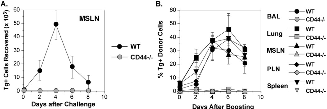
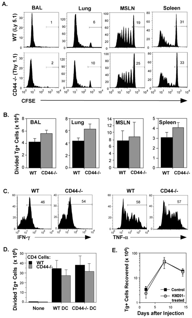
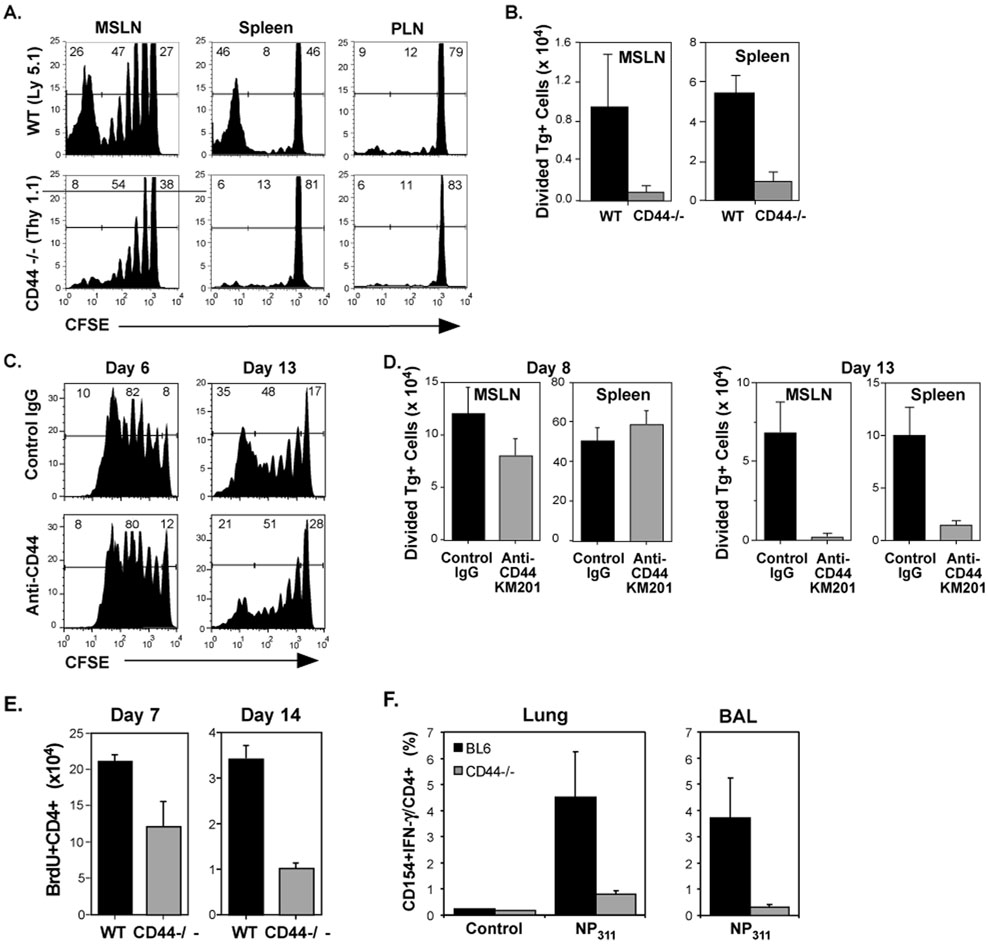
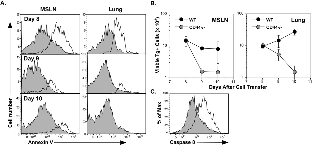
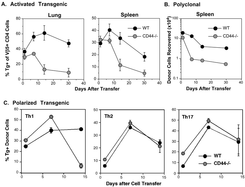
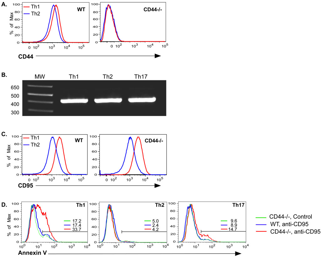
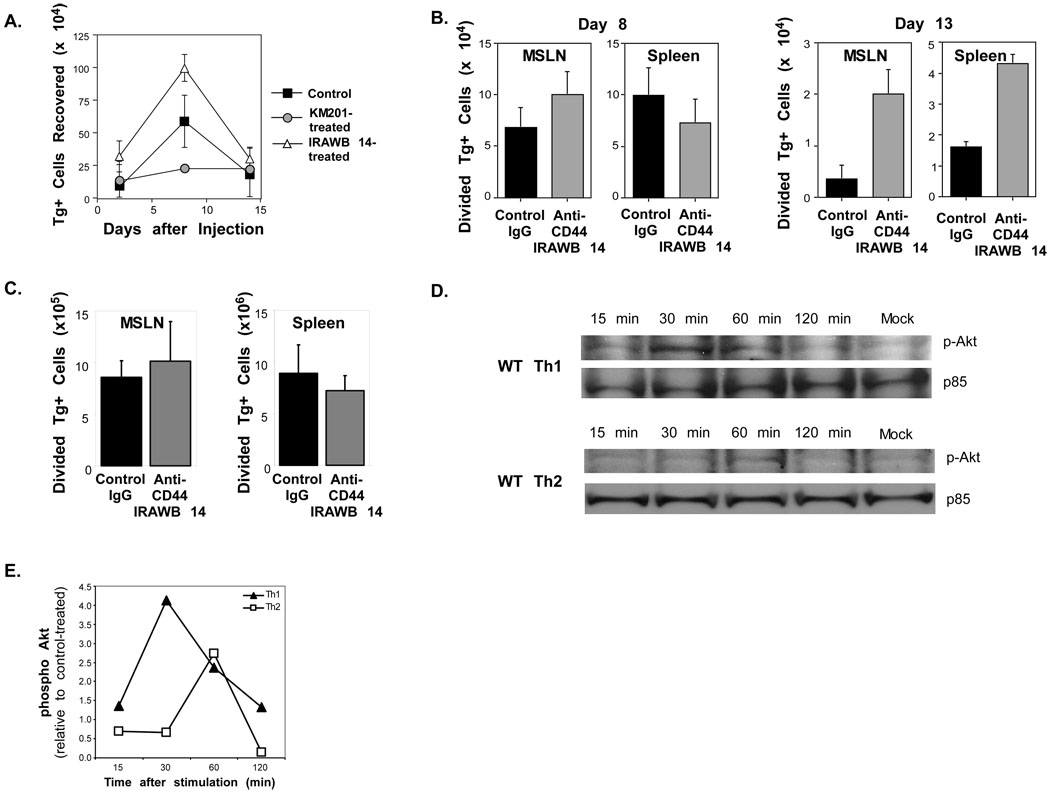
Optimal immunity to microorganisms depends upon the regulated death of clonally expanded effector cells and the survival of a cohort of cells that become memory cells. After activation of naive T cells, CD44, a widely expressed receptor for extracellular matrix components, is upregulated. High expression of CD44 remains on memory cells and despite its wide usage as a "memory marker," its function is unknown. Here we report that CD44 was essential for the generation of memory T helper 1 (Th1) cells by promoting effector cell survival. This dependency was not found in Th2, Th17, or CD8(+) T cells despite similar expression of CD44 and the absence of splice variants in all subsets. CD44 limited Fas-mediated death in Th1 cells and its ligation engaged the phosphoinositide 3-kinase-Akt kinase signaling pathway that regulates cell survival. The difference in CD44-regulated apoptosis resistance in T cell subpopulations has important implications in a broad spectrum of diseases.


Figures

Figure 1
Figure 1. Requirement for CD44 in the generation of memory responses in CD4 cells
CFSE-labeled OT-II cells from WT (Ly5.1) and CD44 −/− (Thy1.1) mice were co-transferred (3×10⁵ each) into C57BL/6 recipients (Ly5.2, Thy1.2) that were then infected with WSN-OVAII. After 22 days, the recipients were challenged with HKx31-OVAII. A) The recovery of Tg+ WT and CD44−/− cells in the MSLN from individual animals. B) The percentage Tg+ cells in the Vβ5+, CD4+ population from BAL, lung, MSLN, PLN and spleen (Mean ± SEM, n = 3–4/group).
Figure 2
Figure 2. CD44-independence of CD4 cell priming
C57BL/6 mice were injected with CFSE-labeled WT and CD44−/− OT-II cells (1.5×10⁶ each) and infected with WSN-OVAII. A) After 8 days, division of Tg+ cells was analyzed by CFSE. The marker on each histogram shows the fraction of undivided cells. B) The average recoveries of Tg+ donor cells that underwent 1 or more divisions on day 8 (Mean ± SEM, n = 3–4/group). C) IFN-γ and TNF-α production by WT and CD44−/− cells after overnight re-stimulation by OVAII peptide with splenic APC. D) CFSE-labeled WT and CD44−/− OT-II cells were co-injected into C57BL/6 recipients as for (A) together with 2×10⁵ CD11c+, OVAII peptide-pulsed DCs from either WT or CD44−/− C57B/6 mice. Recovery of donor CD4 cells that had undergone 1 or more divisions in the spleen 4 days later (Mean ± SEM, n = 4/group). E) WT OT-II Th1 cells were generated with APC and OVAII peptide in the presence of the blocking anti-CD44 mAb, KM201, or control IgG. The cells were then injected into separate groups of C57BL/6 recipients (2×10⁶/mouse). The donor Tg+ cells recovered in the pooled lymph node (LN) and spleens of mice are shown at the indicated times after injection (Mean ± SEM, n = 3–4/group).
Figure 3
Figure 3. Loss of CD4 effectors in the absence of CD44 engagement
C57BL/6 recipients were injected with CFSE-labeled WT and CD44−/− OT-II cells (1.5×10⁶ each) and infected with WSN-OVAII. On day 13 after infection, division (A) and recovery (B) of Tg+ cells was determined as for Figure 2 in the MSLN and spleen. C–D) Recipients of CFSE-labeled WT OT-II cells were injected with either KM201 anti-CD44 mAb or control IgG at the time of cell transfer and infection with WSN-OVAII influenza virus and 3 more times at 3 day intervals. C) The division of the donor cells on days 6 and 13. D) Recovery of donor CD4 cells that had undergone 1 or more divisions in the MSLN and spleen 8 and 13 days after infection. E–F) WT and CD44−/− mice were infected with PR8 influenza virus. E) The mice were treated with BrdU for 7 days before sampling. The recovery of BrdU+ CD4 cells is shown. F) On day 21 after infection, the virus-specific CD4 response was assessed by intracellular staining of cells from the lungs and BALs after overnight culture with anti-CD28 in the presence or absence of NP311 peptide. . Shown are the percentages of CD154+IFN-γ+ virus-specific CD4 cells in the lung. B, D–F, Mean + SEM, n=3–4/group.
Figure 4
Figure 4. Induction of apoptosis in responding CD4 cells deficient in CD44
C57BL/6 recipients were given WT and CD44−/− OT-II cells (1.5×10⁶ each) and infected with WSN-OVAII. A) Apoptosis was assessed by binding of Annexin V and exclusion of 7AAD by WT Tg+ cells (shaded histograms) and CD44−/− Tg+ cells (open histograms) in the indicated tissues. B) Viable recoveries of WT and CD44−/− donor Tg+ cells in the MSLN and lungs (Mean ± SEM, n = 5/group). C) Caspase 8 activation was assessed using a fluorophore-modified substrate with dispersed MSLN cells from recipients of WT and CD44−/− CD4 cells on day 7 after infection. The fluorescence induced by activated caspase 8 for WT Tg+ cells (shaded histogram) and CD44−/− Tg+ cells (open histogram) is shown in 7AAD- Tg+ population. The results are representative of those from 6 recipients.
Figure 5
Figure 5. Impaired survival of activated CD4 in naive recipients in the absence of CD44
A) WT and CD44−/− OT-II cells were stimulated in vitro with APC and OVA peptide and then co-injected (1.5×10⁶ each) into naive C57BL6 recipients. The frequencies of Tg+ cells, gated on the Vβ5+, CD4+ population in the lungs and spleen at the indicated times after cell transfer are shown (Mean ± SEM, n = 4/group). B) Polyclonal, non-Tg WT CD4 cells (Thy1.1, Ly5.2) and CD44−/− CD4 cells (Thy1.2, Ly5.2) were stimulated with anti-CD3/anti-CD28 and co-injected into Ly5.1, Thy1.2 recipients in a dose of 1.5×10⁶/recipient. The recovery of donor cells in the spleen is shown (Mean ± SEM, n = 4/group). C) Th1, Th2, and Th17 cells were generated from OT-II cells with APC and OVAII peptide. Allelically-marked WT and CD44−/− cells of each of the corresponding subsets were co-injected in a dose of 1.5×10⁶ each into C57BL/6 recipients. Shown are the frequencies of donor cells recovered at the indicated times after cell transfer after gating on the Vβ5+, CD4+ population.
Figure 6
Figure 6. CD44s regulates Fas-mediated death in Th1 cells
A) Th1 and Th2 cells were generated from WT and CD44−/− OT-II Thy1.1 cells with OVAII peptide and APC and tested for expression of CD44. B) Th1, Th2, and Th17 cells were generated with WT OT-II cells. RNA was isolated and tested for the presence of CD44 splice variants by RT-PCR using primers for the constant regions that flank the variant region. CD44 standard is 428bp in size. C) Expression of Fas (CD95) on Th1 and Th2 cells. D) Th1, Th2, and Th17 cells were generated from WT and CD44−/− CD4 cells and re-cultured overnight in the presence or absence of plate bound anti-Fas mAb. The number after the colored bars indicate the percentages of cells undergoing apoptosis as indicated by binding of Annexin V and exclusion of 7AAD.
Figure 7
Figure 7. Requirement for CD44 signaling in Th1 CD44−/−
A) WT OT-II Th1 cells were generated with APC and OVAII peptide. The cells were then injected into C57BL/6 recipients (1.5×10⁶/mouse) and treated with control IgG, KM201, or IRAWB 14 on the day of cell transfer, and 3 more times at 3 day intervals. The donor Tg+ cells recovered in the pooled LN and spleens are shown at the indicated times after injection. B) C57BL/6 recipients of 1.5×10⁶ CFSE-labeled WT OT-II cells were injected with either IRAWB 14 or control IgG at the time of cell transfer and infection with WSN-OVAII influenza virus. The antibodies were administered 3 more times at 3-day intervals. The recoveries of donor CD4 cells that had undergone 1 or more divisions in the MSLN and spleen were measured 8 and 13 days later. C). C57BL/6 recipients of 1.5×10⁶ CFSE-labeled WT OT-II cells were injected with either IRAWB 14 or control IgG 8 days after infection with WSN-OVAII influenza virus. The recoveries of donor CD4 cells that had undergone 1 or more divisions in the MSLN and spleen were measured at 10 days after infection. A–C, Mean ± SEM, n = 3–4/group. D, E) Th1 and Th2 cells were generated from WT and CD44−/− C57BL/6 CD4 cells by stimulation with plate-bound anti-CD3 and anti-CD28. After resting for 1 day in rIL-7 and a further day without, the cells were cultured for the indicated times with plate-bound IRAWB 14 mAb. D) Phospho-Akt was detected by western blots and compared to the p85 subunit of PI3K as a loading control. E) Densitometry of phospho Akt on western blot data in C. Results are represented as a ratio between band densities for IRAWB 14- and unstimulated control cells and are corrected for loading differences.

References

    1. Barnden MJ, Allison J, Heath WR, R CF. Defective TCR expression in transgenic mice constructed using cDNA based a- and b- chain genes under the control of heterologous regulatory elements. Immunol. Cell. Biol. 1998;76:34–40. - PubMed
    1. Boyman O, Purton JF, Surh CD, Sprent J. Cytokines and T-cell homeostasis. Curr. Opin. Immunol. 2007;19:320–326. - PubMed
    1. Camp RL, Kraus TA, Pure E. Variations in the cytoskeletal interaction and posttranslational modification of the CD44 homing receptor in macrophages. J Cell Biol. 1991;115:1283–1292. - PMC - PubMed
    1. Chapman TJ, Castrucci MR, Padrick RC, Bradley LM, Topham DJ. Antigen-specific and non-specific CD4+ T cell recruitment and proliferation during influenza infection. Virology. 2005;340:296–306. - PubMed
    1. DeGrendele HC, Kosfiszer M, Estess P, Siegelman MH. CD44 activation and associated primary adhesion is inducible via T cell receptor stimulation. J. Immunol. 1997;159:2549–2553. - PubMed

Publication types

Substances