Review
. 2010 May;1804(5):1041-8.
doi: 10.1016/j.bbapap.2010.01.006. Epub 2010 Jan 15.

The kinetic and chemical mechanism of high-fidelity DNA polymerases

Affiliations
Review

The kinetic and chemical mechanism of high-fidelity DNA polymerases

Kenneth A Johnson. Biochim Biophys Acta. 2010 May.

Abstract

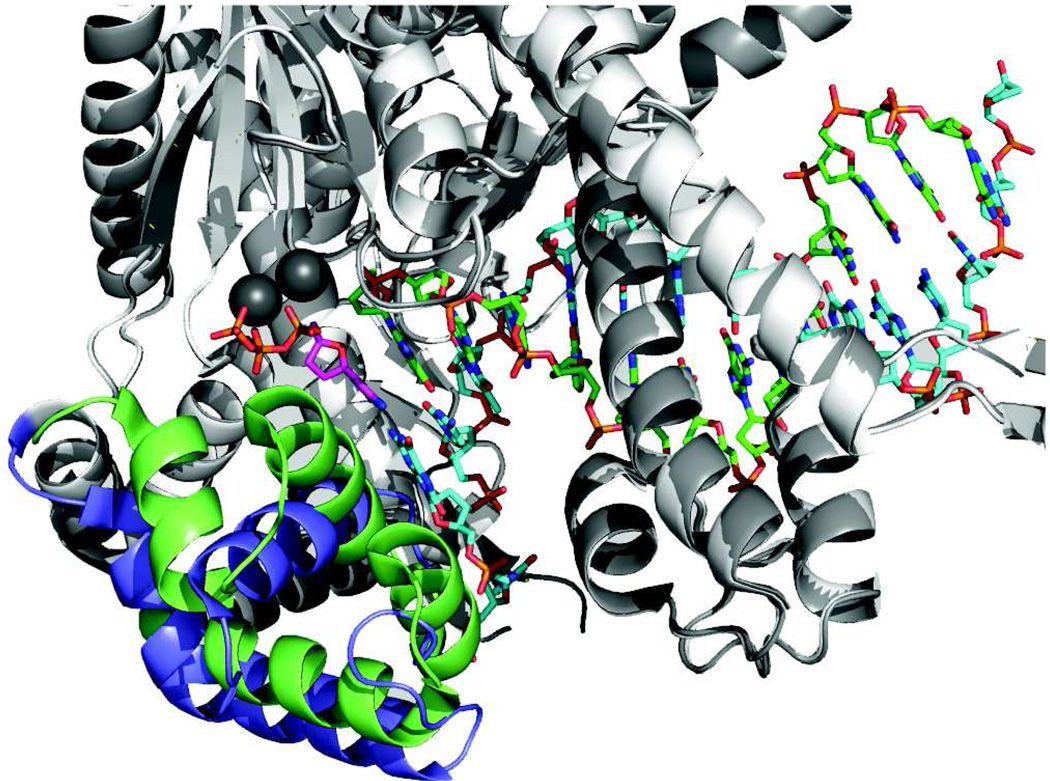
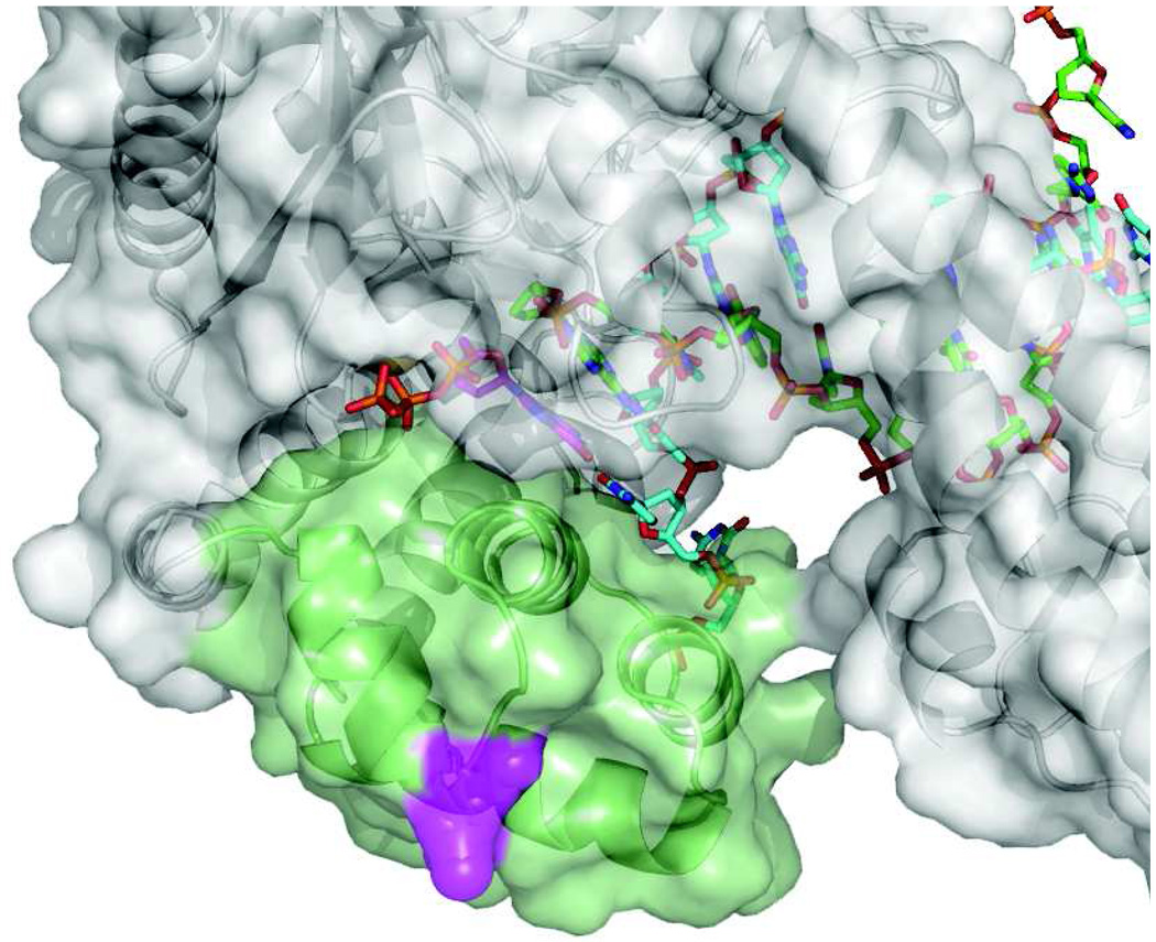
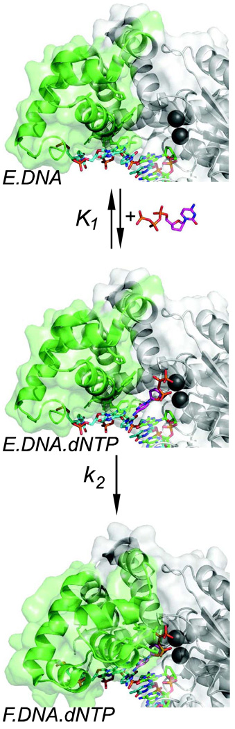
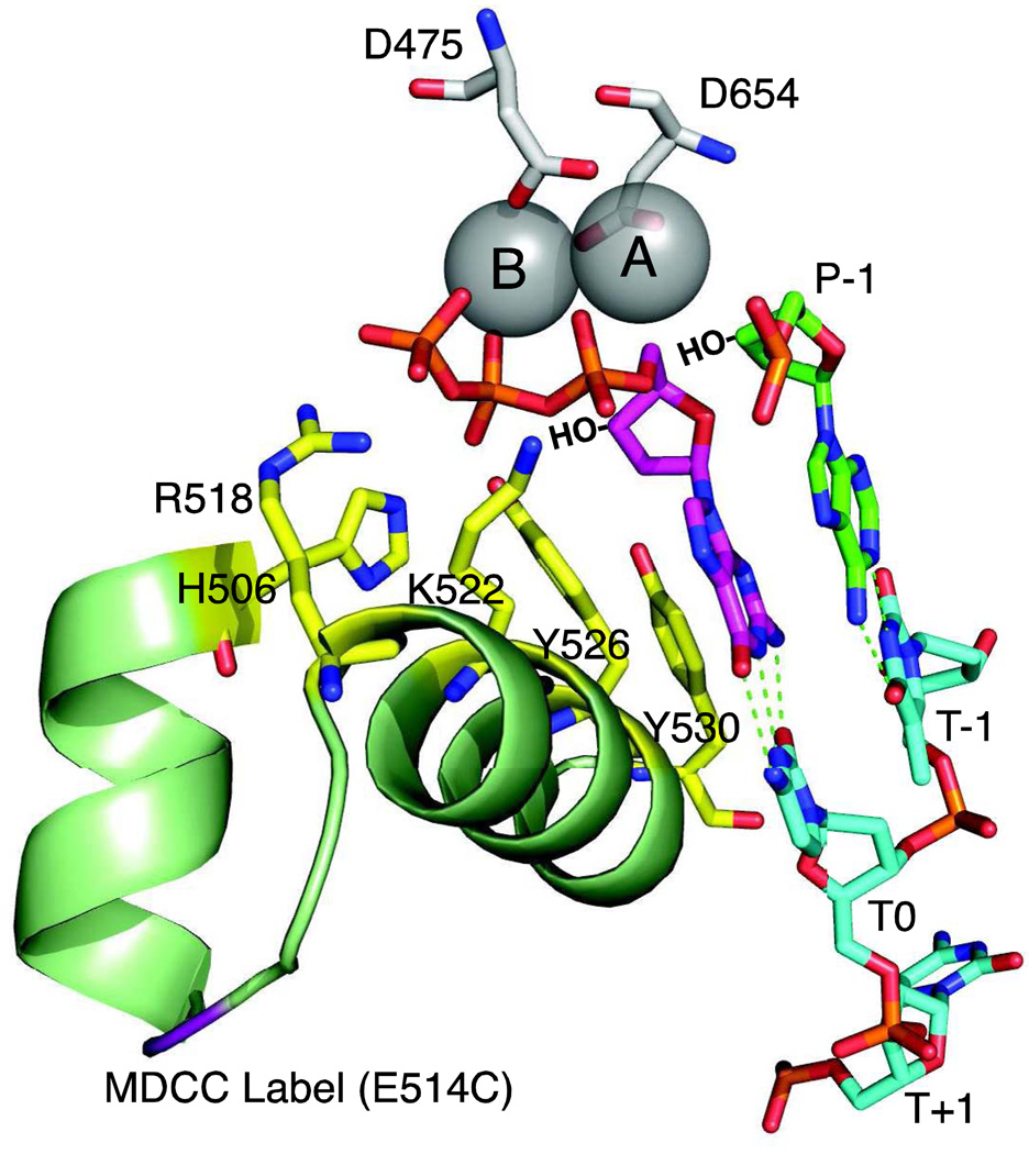
This review summarizes our current understanding of the structural, kinetic and thermodynamic basis for the extraordinary accuracy of high-fidelity DNA polymerases. High-fidelity DNA polymerases, such as the enzyme responsible for the replication of bacteriophage T7 DNA, discriminate against similar substrates with an accuracy that approaches one error in a million base pairs while copying DNA at a rate of approximately 300 base pairs per second. When the polymerase does make an error, it stalls, giving time for the slower proofreading exonuclease to remove the mismatch so that the overall error frequency approaches one in a billion. Structural analysis reveals a large change in conformation after nucleotide binding from an open to a closed state. Kinetic analysis has shown that the substrate-induced structural change plays a key role in the discrimination between correct and incorrect base pairs by governing whether a nucleotide will be retained and incorporated or rapidly released.


Figures

Figure 1
Figure 1. Conformational change upon nucleotide binding
Structures of T7 DNA polymerase in the presence (green and dark gray) and absence (blue and light gray) of nucleotide are aligned to show the changes in structure of the recognition domain (green or blue). Very little change in structure was seen in the remainder of the protein (light or dark gray), which provided a basis for the alignment. The metal ions are dark gray, the incoming nucleotide is magenta, the primer strand is green and the template strand is cyan. The recognition domain is defined as residues N502 to P560. Drawn using Pymol with PDB files 1tk5 (with nucleotide) and 1tk0 (without nucleotide) [62].
Figure 2
Figure 2. Location of the MDCC label
The location of the MDCC label on the surface of the recognition domain is shown in magenta. The recognition domain is green. From 1tk5.pdb.
Figure 3
Figure 3. Pathway of nucleotide binding
The two-step sequence for nucleotide binding is shown with structures of the open E.DNA complex (tko.pdb) and the closed F.DNA.dNTP complex (tk5.pdb). The structure of the open E.DNA.dNTP complex is not known and is modeled here by placing dNTP into the empty E.DNA complex.
Figure 4
Figure 4. Polymerase active site residues
The active site of T7 DNA polymerase is shown derived from 1T7P.pdb [1]. Aspartate residues D475 and D654 hold two metal ions (A and B) in place. Important catalytic residues from the fingers domain, (R518, H506, K522, Y526 and Y530, shown in yellow) make contact only in the closed state. The incoming dGTP is shown in magenta, the primer is in green and the template in cyan. Template positions are labeled T−1 through T+1. The 3'OH groups, lacking in the crystal structure, are shown by HO−. The site of the MDCC label is shown in purple.
Figure 5
Figure 5. Alpha-thio nucleotide
The structure of the dNTP-αS(Sp) is shown with the sulfur represented by the yellow sphere. Also shown is the residue K522 which forms a bond to the Sp oxygen on the α-phosphate and may cause steric effects upon sulfur substitution. The nucleotide is shown in magenta and the metal ions A and B are gray. Drawn from 1T7P.pdb.
Scheme 1
Scheme 1
Scheme 2
Scheme 2
Kinetics of correct nucleotide binding
Scheme 3
Scheme 3
Kinetics of misincorporation

Similar articles

Cited by

References

    1. Doublie S, Tabor S, Long AM, Richardson CC, Ellenberger T. Crystal structure of a bacteriophage T7 DNA replication complex at 2.2 A resolution. Nature. 1998;391:251–258. - PubMed
    1. Huang HF, Chopra R, Verdine GL, Harrison SC. Structure of a covalently trapped catalytic complex of HIV-I reverse transcriptase: Implications for drug resistance. Science. 1998;282:1669–1675. - PubMed
    1. Sarafianos SG, Clark AD, Jr, Tuske S, Squire CJ, Das K, Sheng D, Ilankumaran P, Ramesha AR, Kroth H, Sayer JM, Jerina DM, Boyer PL, Hughes SH, Arnold E. Trapping HIV-1 reverse transcriptase before and after translocation on DNA. J.Biol.Chem. 2003;278:16280–16288. - PubMed
    1. Pelletier H, Sawaya MR, Kumar A, Wilson SH, Kraut J. Structures of Ternary Complexes of Rat Dna-Polymerase-Beta, A Dna Template-Primer, and Ddctp. Science. 1994;264:1891–1903. - PubMed
    1. Li Y, Korolev S, Waksman G. Crystal structures of open and closed forms of binary and ternary complexes of the large fragment of Thermus aquaticus DNA polymerase I: structural basis for nucleotide incorporation. EMBO J. 1998;17:7514–7525. - PMC - PubMed

Publication types

LinkOut - more resources