. 2010 Mar 30;399(1):65-76.
doi: 10.1016/j.virol.2009.12.026. Epub 2010 Jan 15.

Development of quantitative and high-throughput assays of polyomavirus and papillomavirus DNA replication

Affiliations

Development of quantitative and high-throughput assays of polyomavirus and papillomavirus DNA replication

Amélie Fradet-Turcotte et al. Virology. .

Abstract

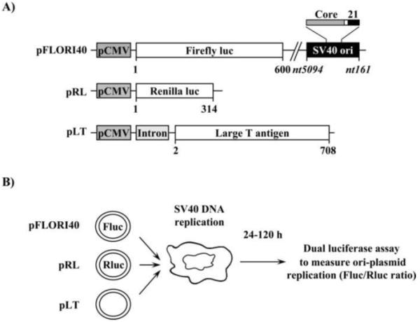
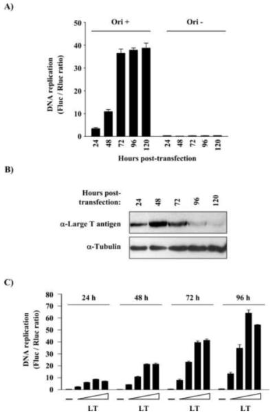
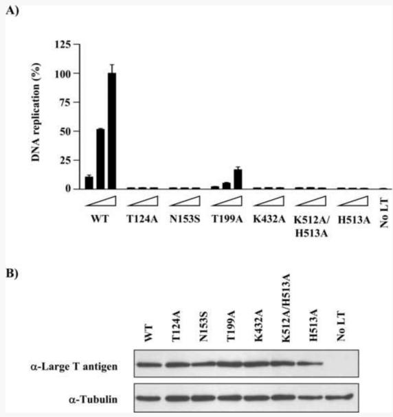
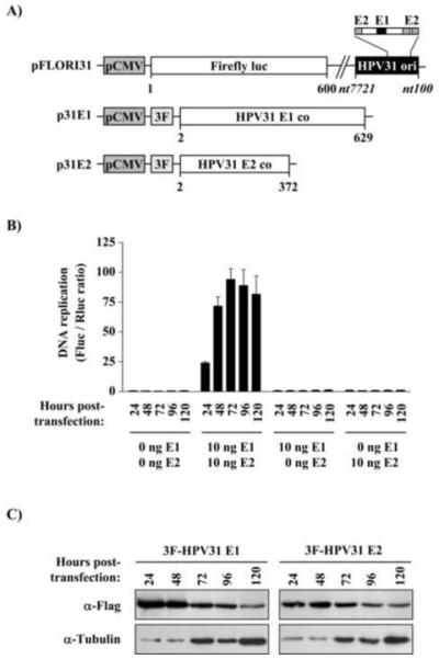
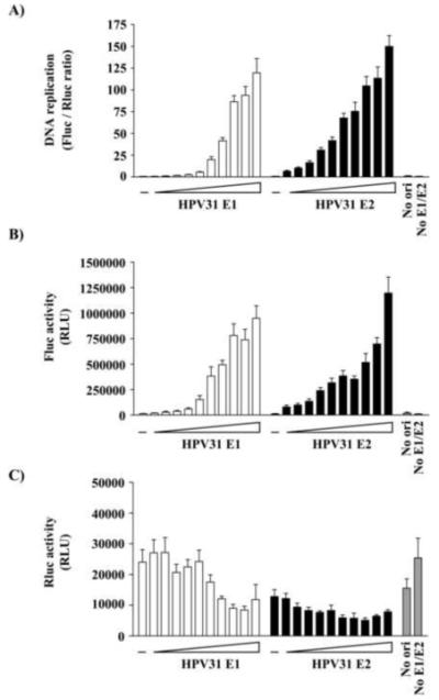
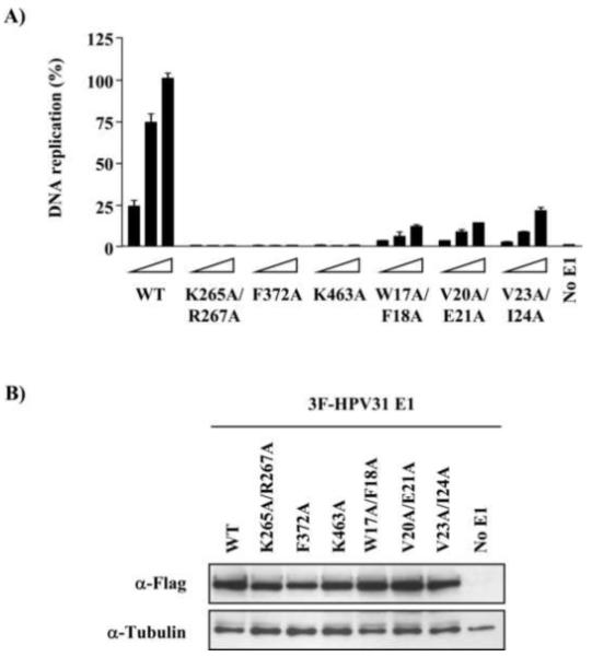
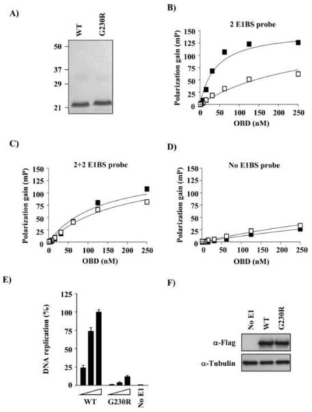
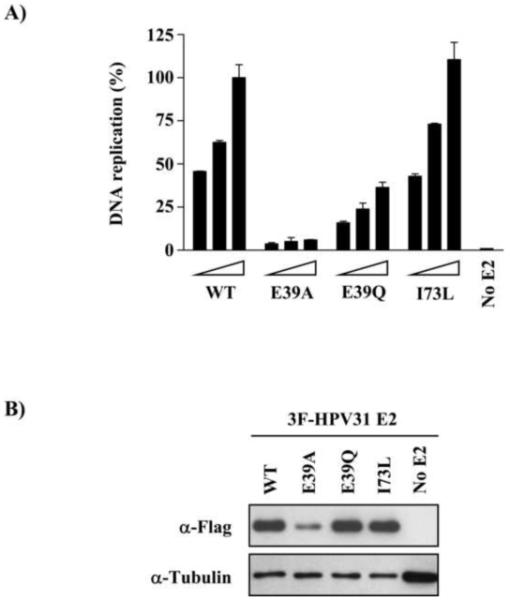
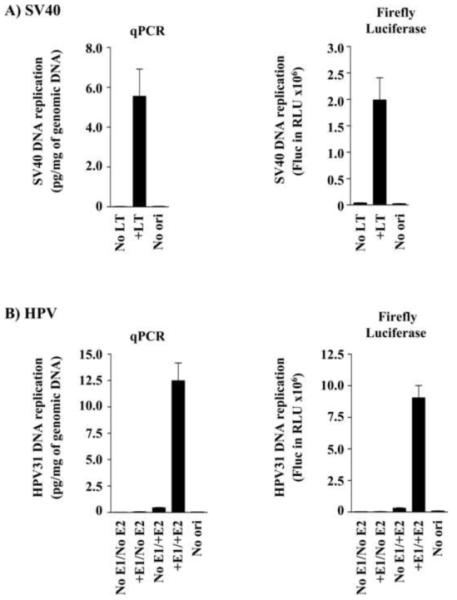
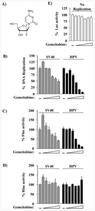
Polyoma- and papillomaviruses genome replication is initiated by the binding of large T antigen (LT) and of E1 and E2, respectively, at the viral origin (ori). Replication of an ori-containing plasmid occurs in cells transiently expressing these viral proteins and is typically quantified by Southern blotting or PCR. To facilitate the study of SV40 and HPV31 DNA replication, we developed cellular assays in which transient replication of the ori-plasmid is quantified using a firefly luciferase gene located in cis to the ori. Under optimized conditions, replication of the SV40 and HPV31 ori-plasmids resulted in a 50- and 150-fold increase in firefly luciferase levels, respectively. These results were validated using replication-defective mutants of LT, E1 and E2 and with inhibitors of DNA replication and cell-cycle progression. These quantitative and high-throughput assays should greatly facilitate the study of SV40 and HPV31 DNA replication and the identification of small-molecule inhibitors of this process.


Figures

Fig. 1
Fig. 1. Principle of the luciferase SV40 DNA replication assay
(A) Schematic representation of the three plasmids used in the assay. The name of each plasmid is written on the left. The location of the SV40 origin of replication is represented by a black box with the position of the core (grey) and 21 bp-repeat regions (black) enlarged above. The nucleotide (nt) sequence boundaries of the origin are indicated. The locations of the CMV promoter and intron are indicated by dark and light grey boxes, respectively. The coding regions of firefly and Renilla luciferase as well as those of LT are indicated by white boxes. Amino acid boundaries of each protein are indicated below each box. (B) Schematic representation of the assay. A plasmid expressing SV40 LT (pLT) is co-transfected in cells along with a second plasmid containing the SV40 origin of replication (pFLORI40) and a firefly luciferase reporter gene. A third plasmid expressing Renilla luciferase (pRL) is also transfected as an internal control to normalize for variations in transfection efficiency. Viral DNA replication is measured using a dual-luciferase assay, at different times post-transfection.
Fig. 2
Fig. 2. Dependence of the SV40 DNA replication assay on time and expression of T antigen
(A) Replication of the SV40 origin-containing plasmid (Ori+), or of a similar plasmid lacking the origin (Ori−), as a function of time post-transfection (in hours). C33A cells were transfected with 12.5 ng of pLT together with 2.5 ng of pFLORI40 and 0.5 ng of RL, and luciferase activities measured at the indicated time points post-transfection. (B) Western blot showing the expression of LT at different time points post-transfection. Tubulin was used as a loading control. Note that the time-dependent increase in tubulin levels reflects the fact that cells continued to proliferate over the course of this 5-day assay. (C) Replication of the SV40 origin-containing plasmid by increasing amounts of pLT (12.5, 25, 50 and 100 ng). Cells transfected without the LT expression vector (−) were used as a negative control. Replication by a gradient of LT was determined at 24, 48, 72 and 96 h, as indicated.
Fig. 3
Fig. 3. Validation of the SV40 DNA replication assay using T antigen mutants
(A) Replication activity of mutant T antigens in C33A cells. Each mutant was tested in three different amounts of pLT (2.5, 6.25 and 12.5 ng). Cells transfected without LT expression vectors (No LT) was used as a negative control. Replication activity is reported as a percentage (%) of the Fluc/Rluc ratio obtained with 12.5 ng of the LT wild-type, which was set at 100. (B) Western blot showing the expression of the different T antigen mutant proteins. Tubulin was used as a loading control.
Fig. 4
Fig. 4. Principle of the luciferase-based HPV31 DNA replication assay
(A) Schematic representation of the three HPV plasmids used in the luciferase assay. The name of each plasmid is written on the left. The location of the CMV promoter and 3F epitope are indicated as dark and light grey boxes, respectively. The coding regions of firefly and Renilla luciferase as well as those of codon-optimized (co) E1 and E2 are indicated by white boxes. Amino acid boundaries are indicated below each box. The location (black box) and nucleotide boundaries of the HPV31 origin of replication are indicated with the position of E1 (black) and E2 (grey) binding sites enlarged above. (B) DNA replication activities measured in C33A cells transfected with the indicated amount of E1 and E2 expression vectors together with 2.5 ng of pFLORI31 and 0.5 ng of pRL. DNA replication activities are expressed as Fluc/Rluc ratios and were determined at 24, 48, 72, 96 and 120 h post-transfection, as indicated. (C) Western blots showing the expression of E1 and E2 at different time points post-transfection. Tubulin was used as a loading control. Note that the time-dependent increase in tubulin levels reflects the fact that cells continued to proliferate over the course of this 5-day assay.
Fig. 5
Fig. 5. Dependence of the HPV31 DNA replication assay on E1 and E2
(A) Replication of the HPV31 origin-containing plasmid in C33A cells transfected with increasing amounts of E1 (white bars) and E2 (black bars) expression vectors (2-fold increments ranging from 0.01 to 25 ng) as described in the text. Cells transfected without the E1 or E2 expression vector (−) were used as negative controls. Cells transfected without E1 and E2 expression vectors (No E1/E2) as well as cells transfected with E1 and E2 but with a plasmid lacking the origin (No ori) were used as controls and are represented as grey bars. (B) and (C) Firefly and Renilla luciferase values (reported as relative light units, RLU) that were used to calculate the ratios presented in (A).
Fig. 6
Fig. 6. Validation of the HPV31 DNA replication assay using E1 mutants
(A) DNA replication activities of the indicated E1 mutants were tested using three different amounts of expression vector (2.5, 5 and 10 ng). Cells transfected without E1 expression vector (No E1) was used as a negative control. Replication activity is reported as a percentage (%) of the Fluc/Rluc ratio obtained with 10 ng of the E1 wild-type. (B) Anti-Flag Western blots showing the expression of E1 mutant proteins. Tubulin was used as a loading control.
Fig. 7
Fig. 7. Characterization of a dimerization-defective HPV31 E1 mutant
(A) Coomassie-stained SDS-PAGE showing the purified wild-type and G230R E1 OBDs. 3 μg of each protein was loaded on the gel. (B-D) Fluorescence polarization DNA binding assays. Binding isotherms were performed with increasing concentrations of wild-type (filled squares) or G230R (open squares) OBD and 10 nM of fluorescent DNA probe either lacking (No E1BS probe (D)) or containing two E1 binding sites spaced by 3 bp (2 E1BS probe (B)) or 5 bp (2+2 E1BS probe (C)). Only a spacing of 3 bp allows dimerization of the OBD. Each binding isotherm was performed in triplicate. (E) DNA replication activities of wild-type and G230R E1 were tested using three different amounts of expression vector, as described in the text. (F) Anti-Flag Western blots showing the expression of E1 proteins. Tubulin was used as a loading control.
Fig. 8
Fig. 8. Validation of the HPV31 DNA replication assay using E2 mutants
(A) DNA replication activities of the indicated E2 mutants were tested using three different amounts of expression vector (1, 2.5 and 5 ng). Cells transfected without E2 expression vector (No E2) was used as a negative control. Replication activity is reported as a percentage (%) of the Fluc/Rluc ratio obtained with 5 ng of E2 wild-type. (B) Anti-Flag Western blots showing the expression of E2 mutant proteins. Tubulin was used as a loading control.
Fig. 9
Fig. 9. Correlation between ori-plasmid replication and firefly luciferase expression
(A) Levels of SV40 plasmid replication measured by quantitative real-time PCR (qPCR) or determined by measuring levels of firefly luciferase activity. C33A cells were transfected with the pFLORI40 plasmid and the LT expression vector (+LT). As controls, cells were transfected without the LT expression vector (No LT) or with a similar plasmid lacking the ori (No ori). The amounts of DNA replication measured by qPCR are reported in pg of DpnI-resistant plasmid per mg of total genomic DNA. Firefly luciferase (Fluc) activity is reported in relative light units (RLU). (B) A similar analysis was performed for HPV31 DNA replication with the exception that cells were transfected with the pFLORI31 plasmid, or a similar plasmid lacking the ori (No ori), and with expression vectors for E1 and E2, as indicated.
Fig. 10
Fig. 10. Validation of the SV40 and HPV31 assays using the DNA replication inhibitor gemcitabine
(A) Structure of gemcitabine. (B) Replication activity measured in cells treated with increasing concentrations of gemcitabine (2-fold increments ranging from 1.56 to 50 nM) or treated with DMSO as a vehicle control (−). Replication activity is reported as a percentage (%) of the Fluc/Rluc ratio obtained in presence of DMSO only. (C) and (D) Firefly and Renilla luciferase values, reported as a percentage of the values obtained without gemcitabine (DMSO only), that were used to calculate the replication activity levels shown in (B). (E) Effect of gemcitabine on expression of CMV-Fluc in the absence of plasmid replication (No replication) is reported as a percentage (%) of the value obtained in absence of gemcitabine (DMSO only).

Similar articles

Cited by

References

    1. Abbate EA, Berger JM, Botchan MR. The X-ray structure of the papillomavirus helicase in complex with its molecular matchmaker E2. Genes Dev. 2004;18(16):1981–1996. - PMC - PubMed
    1. Androphy EJ, Lowy DR, Schiller JT. Bovine papillomavirus E2 trans-activating gene product binds to specific sites in papillomavirus DNA. Nature. 1987;325(6099):70–3. - PubMed
    1. Auster AS, Joshua-Tor L. The DNA-binding domain of human papillomavirus type 18 E1. Crystal structure, dimerization, and DNA binding. J.Biol.Chem. 2004;279(5):3733–3742. - PubMed
    1. Baxter MK, McPhillips MG, Ozato K, McBride AA. The mitotic chromosome binding activity of the papillomavirus E2 protein correlates with interaction with the cellular chromosomal protein, Brd4. J Virol. 2005;79(8):4806–18. - PMC - PubMed
    1. Bedin M, Gaben AM, Saucier C, Mester J. Geldanamycin, an inhibitor of the chaperone activity of HSP90, induces MAPK-independent cell cycle arrest. Int J Cancer. 2004;109(5):643–52. - PubMed

Publication types

MeSH terms