. 2010 Mar 30;399(1):11-22.
doi: 10.1016/j.virol.2009.12.011. Epub 2010 Jan 18.

Exacerbation of corneal scarring in HSV-1 gK-immunized mice correlates with elevation of CD8+CD25+ T cells in corneas of ocularly infected mice

Affiliations

Exacerbation of corneal scarring in HSV-1 gK-immunized mice correlates with elevation of CD8+CD25+ T cells in corneas of ocularly infected mice

Sariah J Allen et al. Virology. .

Abstract

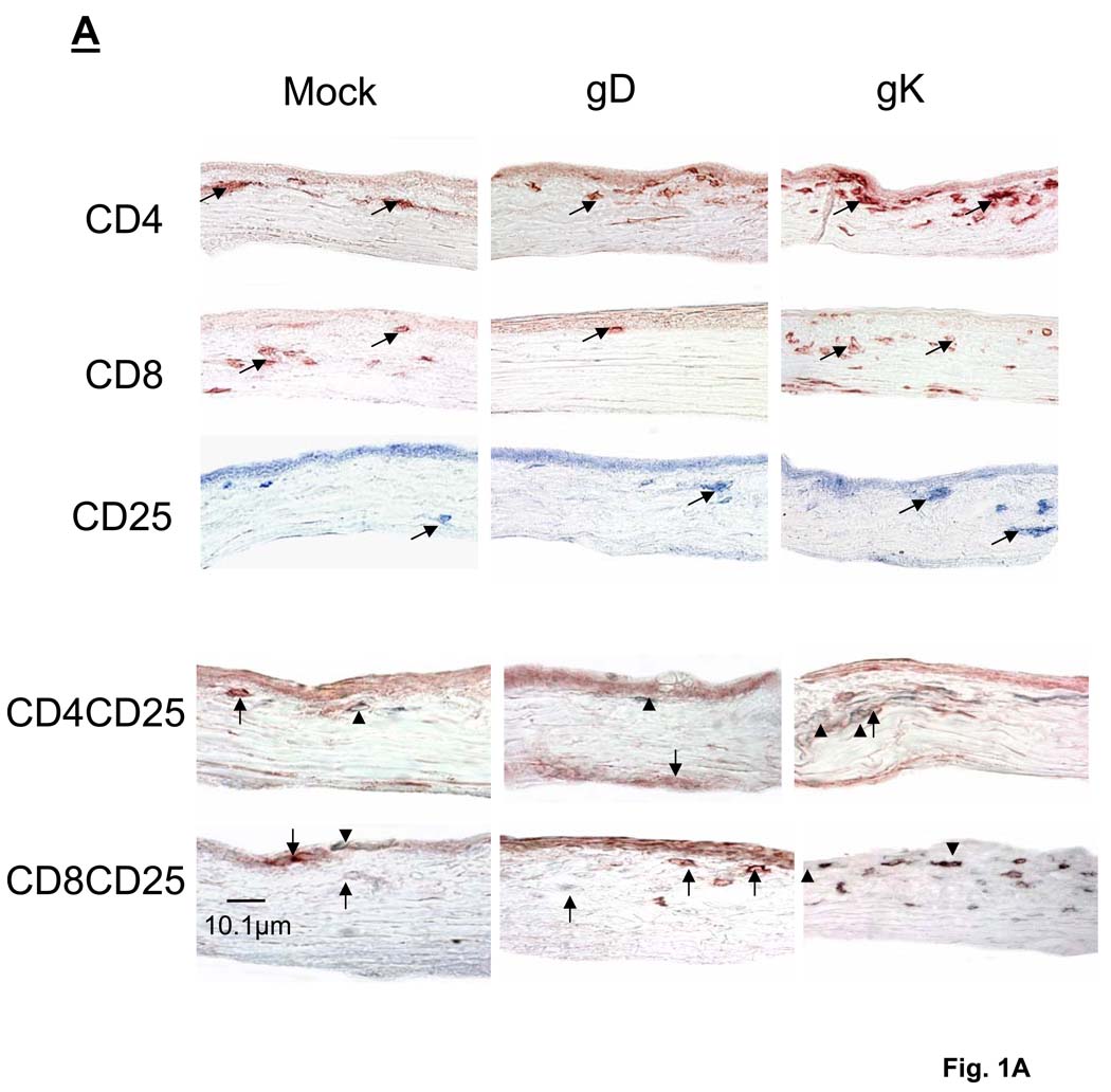
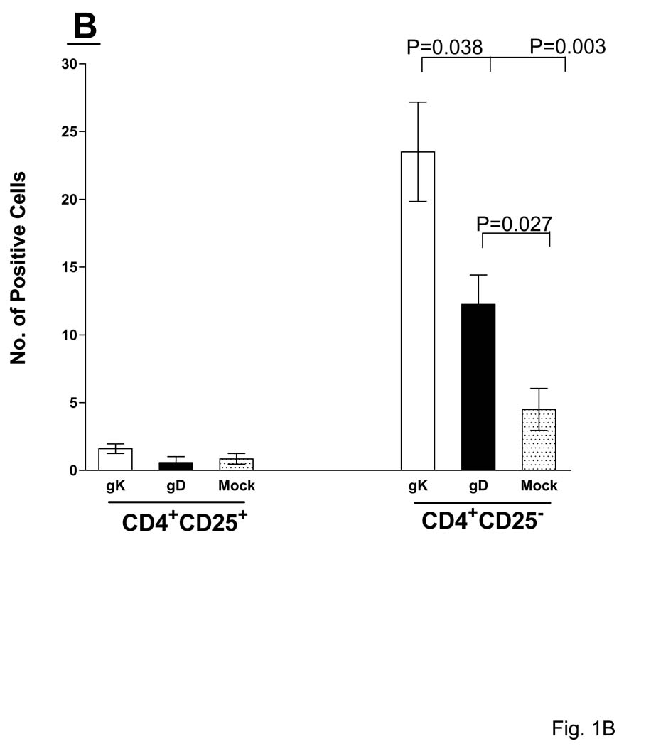
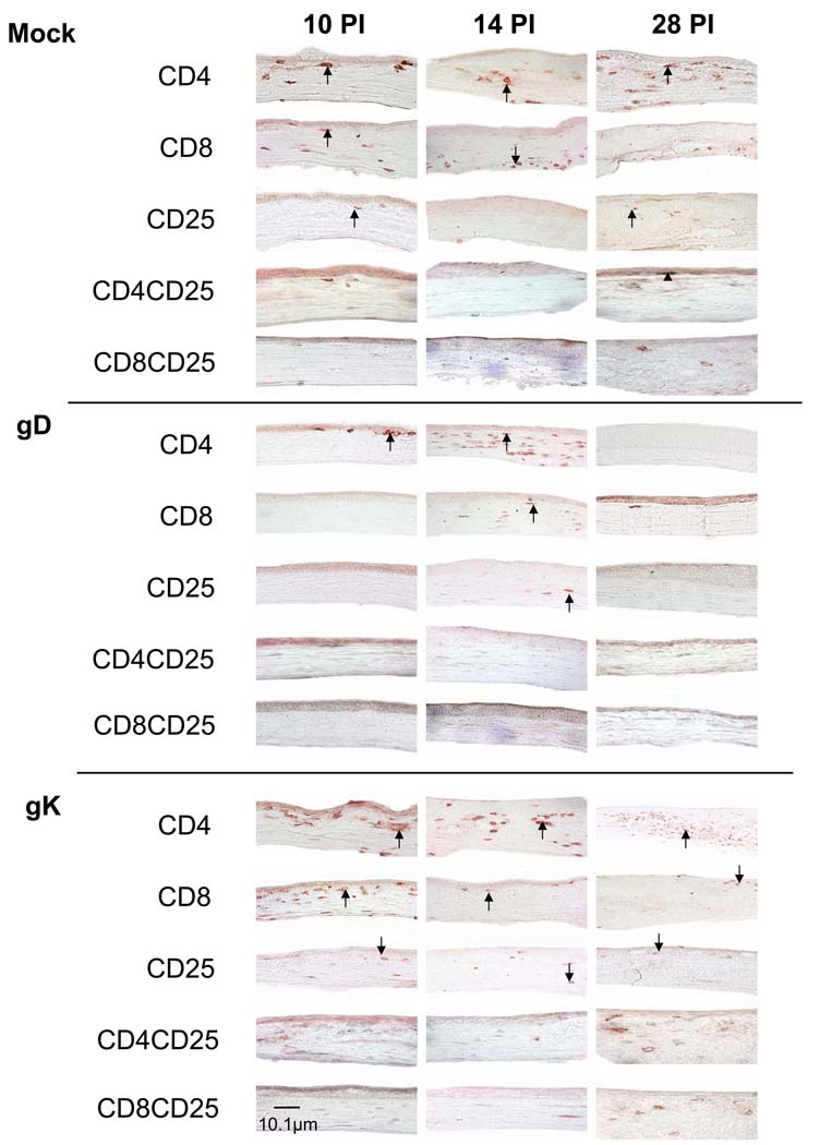
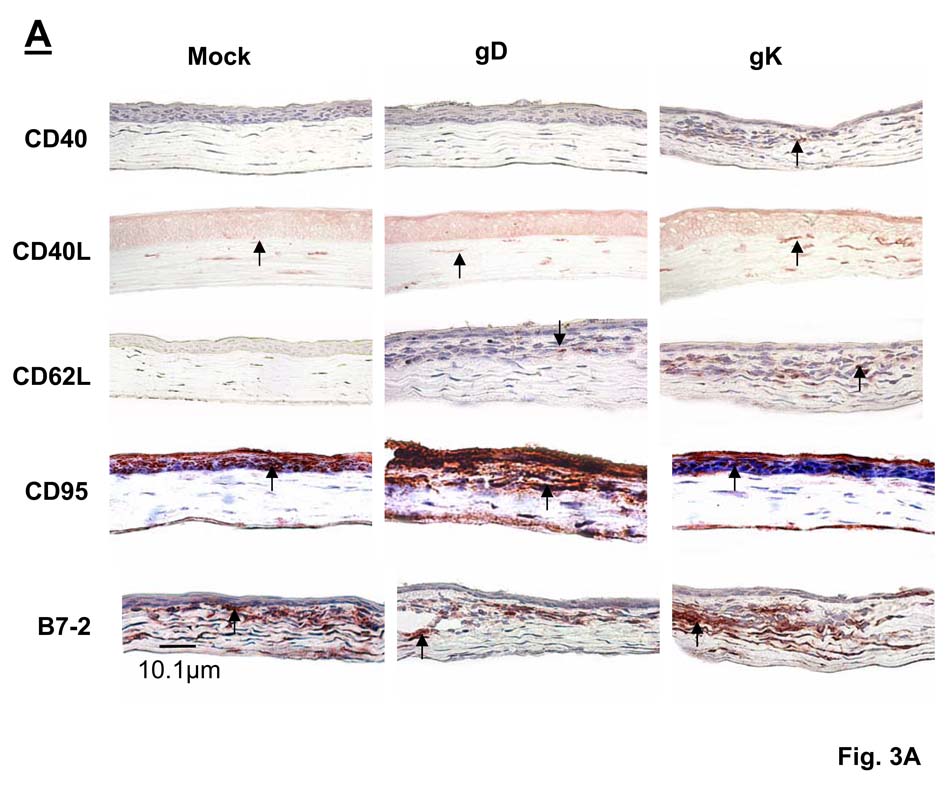
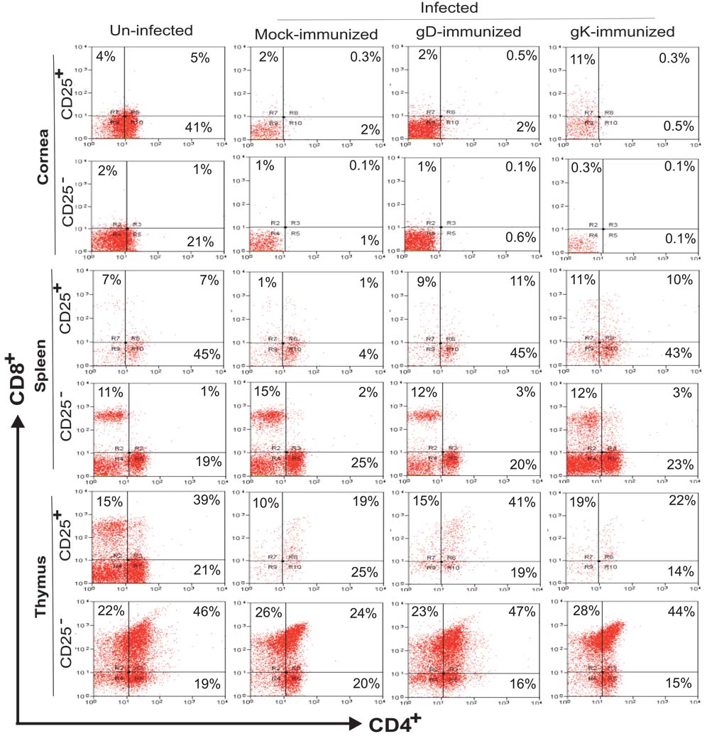
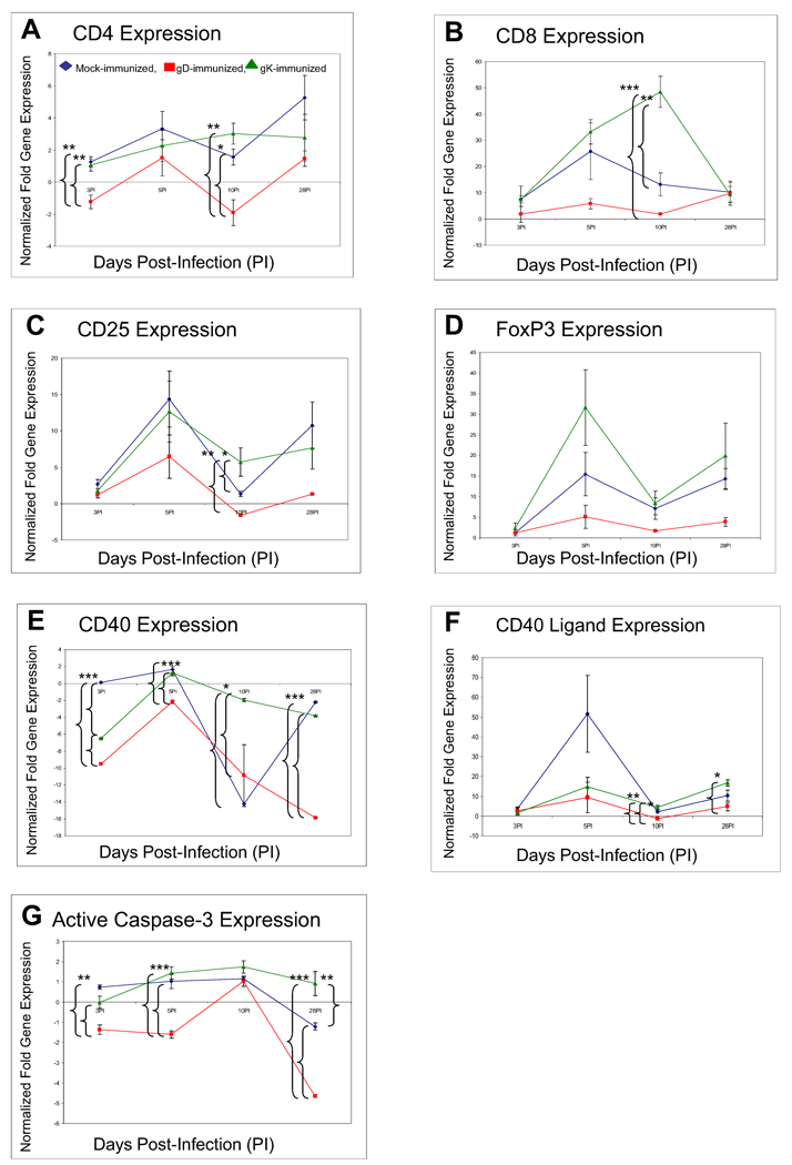
We have shown previously that exacerbation of corneal scarring (CS) in HSV-1 glycoprotein K (gK) immunized mice was associated with CD8+ T cells. In this study, we investigated the type and the nature of the immune responses that are involved in the exacerbation of CS in gK-immunized animals. BALB/c mice were vaccinated with baculovirus expressed gK, gD, or mock-immunized. Twenty-one days after the third immunization, mice were ocularly infected with 2 x 10(5) PFU/eye of virulent HSV-1 strain McKrae. Infiltration of the cornea by CD4+, CD8+, CD25+, CD4+CD25+, CD8+CD25+, CD19+, CD40+, CD40L+, CD62L+, CD95+, B7-1+, B7-2+, MHC-I+, and MHC-II+ cells was monitored by immunohistochemistry, qRT-PCR and FACS at various times post-infection (PI). This study demonstrated for the first time that the presence of CD8+CD25+ T cells in the cornea is correlated with exacerbation of CS in the gK-immunized group.


Figures

Fig. 1
Fig. 1. Immunohistochemistry of T cell infiltrates in corneas of immunized mice on day 5 PI
Mice were immunized and ocularly infected as described in Materials and Methods. Corneas from infected mice were removed at necropsy on day 5 PI, frozen, sectioned, then single or sequential double stained as described in Materials and Methods. (A) T cell infiltrates in cornea. Panels: Red: CD4+ or CD8+; Light blue:CD25+; and Dark-gray: CD4+CD25+ or CD8+CD25+ infiltrates. 60× direct magnification of single stain and 80× magnification of double stain. Arrows point to single stained cells while arrowheads indicate positive double staining. (B) Number of CD4+CD25+ infiltrates in cornea. For each double staining, the number of inflammatory CD4+CD25+ and CD4+CD25 infiltrates on the entire corneal section was counted double-blind for each of six eyes as 1B (above). (C) Number of CD8+CD25+ infiltrates in cornea. For each double staining, the number of inflammatory CD8+CD25+ and CD8+CD25 infiltrates on the entire corneal section was counted double-blind for each of six eyes. Each section represents the width and height of the entire cornea. The height of each bar represents the average number of cellular infiltrates from six eyes ± SEM.
Fig. 2
Fig. 2. Immunohistochemistry of T cell infiltrates in corneas of immunized mice on days 10, 14, and 28 PI
BALB/c mice were immunized and infected as described in the legend to Fig. 1. Corneas from infected mice were removed at necropsy on days 10, 14 and 28 PI, frozen, sectioned, then single CD4+, CD8+, or CD25+ staining or sequential double staining for CD4+CD25+ or CD8+CD25+ on corneal sections was performed as described in Materials and Methods. Individual positive staining for CD4+, CD8+ and CD25+ appears dark red, CD25+ appears light blue/gray, while double staining CD4+CD25+ or CD8+CD25+ appears dark gray due to the cell having both a dark red and light blue label. 60× direct magnification. Arrows indicate positive single staining while arrowheads indicate positive double staining.
Fig. 3
Fig. 3. Immunohistochemistry of various corneal infiltrates in immunized mice on day 5 PI
(A) Corneal Infiltrates. BALB/c mice were immunized and infected as described in the legend to Fig. 1. Corneas were harvested on day 5 PI, snap frozen, and processed as described in Materials and Methods. Representative corneal sections stained with anti-B7-1, anti-B7-2, anti-CD19, anti-CD40, anti-CD40L, anti-CD62L, anti-CD95, anti-MHC-I, or anti-MHC-II are shown. Positive staining appears red, whereas tissue is counterstained blue. 60× direct magnification. (B). Quantification of corneal infiltrates. For each staining, the number of infiltrates on the entire corneal section was counted double-blind for each of six eyes. Each section represents the width and height of the entire cornea. The height of each bar represents the average number of cellular infiltrates from six eyes ± SEM.
Fig. 4
Fig. 4. Detection of CD8+CD25+ T cells in corneas of gK immunized mice by FACS
BALB/c mice were immunized and infected as described in the legend to Fig. 1. Corneas, spleens, and thymuses were harvested on day 5 PI. The corneal tissues were cut into small pieces and then treated with 3 mg/mL of collagenase type I for 2.5 h at 37°C, with gentle passage 3–4 times through an eighteen-gauge syringe after the first hour. The single cell suspensions of total corneal cells, spleens, and thymuses were prepared as described (Osorio et al., 2005). Cell surface staining of single cell suspensions of cornea, spleen, and thymus from individual mice was accomplished using anti-CD4-Cy, anti-CD8a-PE, and anti-CD25-FITC mAbs. Three-color FACS analyses of isolated corneal cells were performed using a FACScan. CD4+ and CD8+ T cells were gated for presence or absence of CD25 expression. Experiments were repeated twice.
Fig. 5
Fig. 5. qRT-PCR analyses of various inflammatory transcripts in corneas of gK-immunized mice at different times PI
BALB/c mice were immunized and infected as described in the legend to Fig. 1. Total RNA from each mouse cornea was isolated on days 3, 5, 10, and 28 PI. CD4, CD8, CD25, CD40, CD40L, active Caspase-3, and FoxP3 expression in naive mice was used to estimate the relative expression of each transcript in corneas of mice in gK-, gD-, or mock-immunized group. GAPDH expression was used to normalize the relative expression of each transcript in corneas of gK-, gD-, or mock-immunized mice. Each point represents the mean ± SEM from 3 mice. Panels: A) CD4 transcript; B) CD8 transcript; C) FoxP3 transcript; D) CD25 transcript; E) CD40 transcript; F) CD40L transcript; and G) active Caspase-3 transcript. *, **, *** significantly different at P<0.05, P<0.01, and P<0.001, respectively.

Similar articles

Cited by

References

    1. Arbones ML, Ord DC, Ley K, Ratech H, Maynard-Curry C, Otten G, Capon DJ, Tedder TF. Lymphocyte homing and leukocyte rolling and migration are impaired in L-selectin-deficient mice. Immunity. 1994;1(4):247–260. - PubMed
    1. Bagenstose LM, Agarwal RK, Silver PB, Harlan DM, Hoffmann SC, Kampen RL, Chan CC, Caspi RR. Disruption of CD40/CD40-ligand interactions in a retinal autoimmunity model results in protection without tolerance. J Immunol. 2005;175(1):124–130. - PubMed
    1. Banerjee K, Deshpande S, Zheng M, Kumaraguru U, Schoenberger SP, Rouse BT. Herpetic stromal keratitis in the absence of viral antigen recognition. Cell Immunol. 2002;219(2):108–118. - PubMed
    1. Barron BA, Gee L, Hauck WW, Kurinij N, Dawson CR, Jones DB, Wilhelmus KR, Kaufman HE, Sugar J, Hyndiuk RA, et al. Herpetic Eye Disease Study. A controlled trial of oral acyclovir for herpes simplex stromal keratitis. Ophthalmology. 1994;101(12):1871–1882. - PubMed
    1. Brandt CR. The role of viral and host genes in corneal infection with herpes simplex virus type 1. Exp Eye Res. 2005;80(5):607–621. - PubMed

Publication types

MeSH terms

Substances