Evaluation of peripheral atherosclerosis: a comparative analysis of angiography and intravascular ultrasound imaging
- PMID: 20080002
- PMCID: PMC2847042
- DOI: 10.1016/j.jvs.2009.11.034
Evaluation of peripheral atherosclerosis: a comparative analysis of angiography and intravascular ultrasound imaging
Abstract
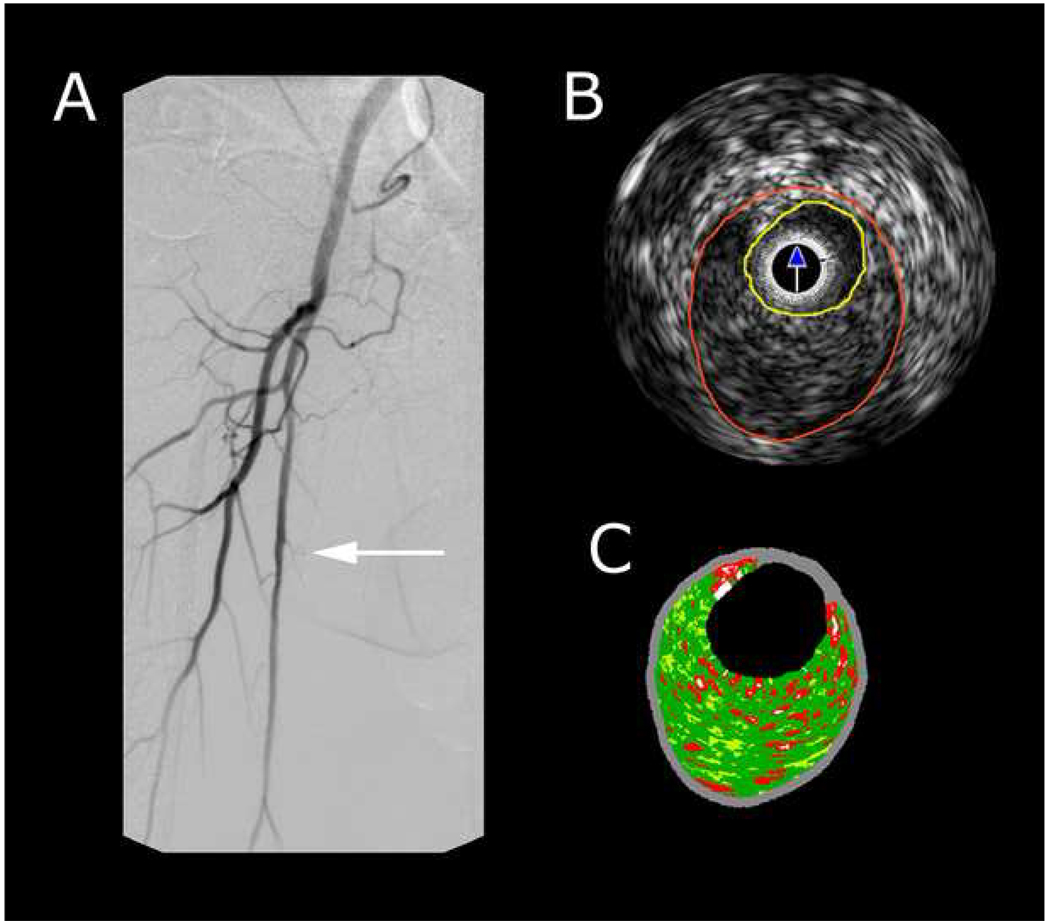
Objective: Angiography remains a critical component for diagnostic imaging and therapeutic intervention in peripheral arterial disease (PAD). The goal of this study was to compare angiography with corresponding intravascular ultrasound (IVUS) imaging of the same vessels in patients with PAD.
Methods: From 2004 to 2008, 93 patients undergoing angiography for PAD were recruited in a prospective observational analysis. At the time of angiography, diseased lower extremities were interrogated using a 10-cm IVUS pullback with registration points. IVUS data were analyzed with radiofrequency techniques for vessel and lumen diameter, plaque volume, plaque composition, and cross-sectional area. Similarly, three vascular surgeons blinded to the IVUS data graded corresponding angiographic images according to vessel diameter, degree of stenosis, degree of calcification, and extent of eccentricity. Statistical analyses of matched IVUS images and angiograms were performed.
Results: The distribution of demographic and risk variables were typical for PAD: 54% male, 96% hypertension, 78% hyperlipidemia, 44% diabetic, 87% tobacco history, 65% coronary artery disease, and 10% end-stage renal disease. Symptoms precipitating the angiographic evaluation included claudication (53%), rest pain (18%), and tissue loss (29%). Angiographic and IVUS interpretation were similar for luminal diameters, but external vessel diameter was greater by IVUS imaging (7.0 +/- 0.7 vs 5.2 +/- 0.8 mm, P < .05). The two-dimensional diameter method resulted in a significant correlation for stenosis determination (r = 0.84); however, IVUS determination of vessel area stenosis was greater by 10% (95% confidence interval, 0.3%-21%, P < .05). IVUS imaging indicated that a higher proportion of plaques were concentric. Grading of calcification was moderate to severe in 40% by angiography but in only 7% by IVUS (P < .05).
Conclusions: In the evaluation of PAD, angiography and IVUS imaging provide similar luminal diameters and diameter-reducing stenosis measurements. Determination of overall vessel diameter and interpretation of plaque morphology by angiography are discordant from IVUS-derived data.
Figures


Similar articles
-
Automatic quantification and characterization of coronary atherosclerosis with computed tomography coronary angiography: cross-correlation with intravascular ultrasound virtual histology.Int J Cardiovasc Imaging. 2013 Jun;29(5):1177-90. doi: 10.1007/s10554-013-0194-x. Epub 2013 Feb 16. Int J Cardiovasc Imaging. 2013. PMID: 23417447
-
Prospective validation of standardized, 3-dimensional, quantitative coronary computed tomographic plaque measurements using radiofrequency backscatter intravascular ultrasound as reference standard in intermediate coronary arterial lesions: results from the ATLANTA (assessment of tissue characteristics, lesion morphology, and hemodynamics by angiography with fractional flow reserve, intravascular ultrasound and virtual histology, and noninvasive computed tomography in atherosclerotic plaques) I study.JACC Cardiovasc Interv. 2011 Feb;4(2):198-208. doi: 10.1016/j.jcin.2010.10.008. JACC Cardiovasc Interv. 2011. PMID: 21349459
-
Intravascular Ultrasound Imaging Versus Digital Subtraction Angiography in Patients with Peripheral Vascular Disease.J Invasive Cardiol. 2020 Mar;32(3):99-103. doi: 10.25270/jic/19.00418. J Invasive Cardiol. 2020. PMID: 32123141
-
Coronary CT angiography versus intravascular ultrasound for estimation of coronary stenosis and atherosclerotic plaque burden: a meta-analysis.J Cardiovasc Comput Tomogr. 2013 Jul-Aug;7(4):256-66. doi: 10.1016/j.jcct.2013.08.006. Epub 2013 Aug 23. J Cardiovasc Comput Tomogr. 2013. PMID: 24148779 Review.
-
[New methods of coronary imaging II. Intracoronary ultrasonography in clinical practice].Ital Heart J Suppl. 2001 Jun;2(6):579-92. Ital Heart J Suppl. 2001. PMID: 11460831 Review. Italian.
Cited by
-
Advanced imaging in limb salvage.Methodist Debakey Cardiovasc J. 2012 Oct-Dec;8(4):28-32. doi: 10.14797/mdcj-8-4-28. Methodist Debakey Cardiovasc J. 2012. PMID: 23342185 Free PMC article. Review.
-
Time-Frequency Analysis of EEG Signals and GLCM Features for Depth of Anesthesia Monitoring.Comput Intell Neurosci. 2021 Aug 11;2021:8430565. doi: 10.1155/2021/8430565. eCollection 2021. Comput Intell Neurosci. 2021. PMID: 34422035 Free PMC article.
-
Intravascular Ultrasound Use in Peripheral Arterial and Deep Venous Interventions: Multidisciplinary Expert Opinion From SCAI/AVF/AVLS/SIR/SVM/SVS.J Soc Cardiovasc Angiogr Interv. 2024 Jan 9;3(1):101205. doi: 10.1016/j.jscai.2023.101205. eCollection 2024 Jan. J Soc Cardiovasc Angiogr Interv. 2024. PMID: 39131984 Free PMC article.
-
Intravascular ultrasound guidance for lower extremity arterial and venous interventions.EuroIntervention. 2022 Sep 20;18(7):598-608. doi: 10.4244/EIJ-D-21-00898. EuroIntervention. 2022. PMID: 35438078 Free PMC article. Review.
-
Intravascular Ultrasound-Guided Femoropopliteal Endovascular Intervention: Could it Become a Standard Procedure?J Atheroscler Thromb. 2023 Sep 1;30(9):1111-1112. doi: 10.5551/jat.ED233. Epub 2023 Apr 26. J Atheroscler Thromb. 2023. PMID: 37100598 Free PMC article. No abstract available.
References
-
- Kruger RA, Mistretta CA, Crummy AB, Sackett JF, Goodsitt MM, Riederer SJ, et al. Digital K-edge subtraction radiography. Radiology. 1977;125(1):243–245. - PubMed
-
- Sones FM., Jr Cine-coronary arteriography. Ohio Med. 1962;58:1018–1019. - PubMed
-
- Vlodaver Z, Frech R, Van Tassel RA, Edwards JE. Correlation of the antemortem coronary arteriogram and the postmortem specimen. Circulation. 1973;47(1):162–169. - PubMed