. 2010 Mar;79(2):93-101.
doi: 10.1016/j.mvr.2010.01.006. Epub 2010 Jan 18.

Chronic whole-body hypoxia induces intussusceptive angiogenesis and microvascular remodeling in the mouse retina

Affiliations

Chronic whole-body hypoxia induces intussusceptive angiogenesis and microvascular remodeling in the mouse retina

Alyssa C Taylor et al. Microvasc Res. 2010 Mar.

Abstract

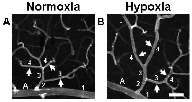
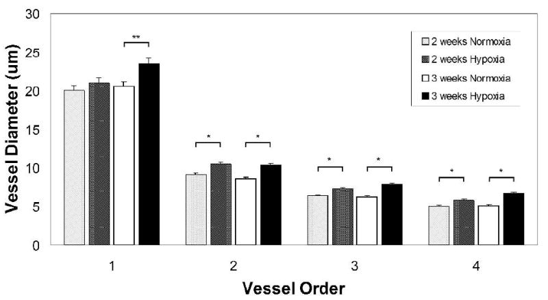
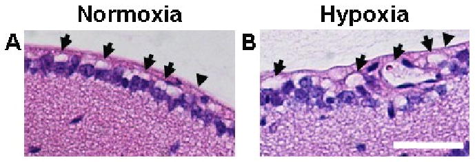
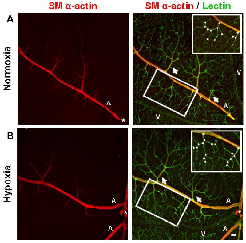
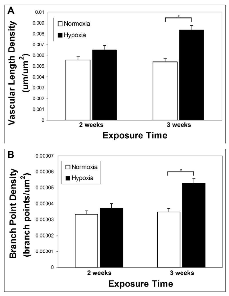
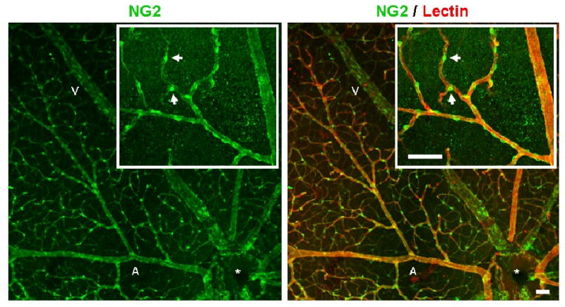
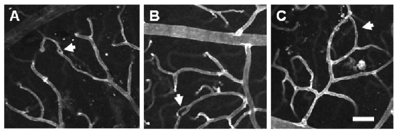
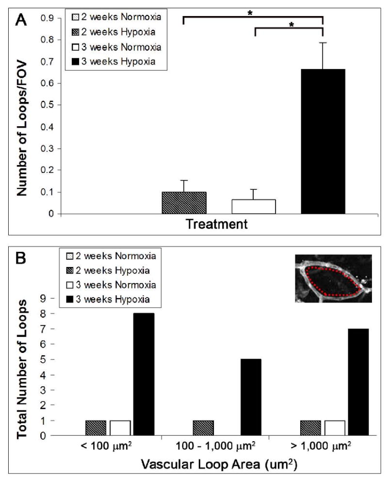
Currently, little is known about the response of the adult retinal microvasculature to hypoxia. To test the hypothesis that chronic systemic hypoxia induces angiogenesis and microvascular remodeling in the adult mouse retina, adult 10-week old female C57Bl/6 mice were exposed to 10% O(2) for 2 or 3 weeks. After hypoxia exposure, retinas were harvested, whole-mounted, and processed for immunohistochemistry. Retinas were stained with lectin, anti-smooth muscle alpha-actin antibody, and anti-NG2 antibody to visualize microvascular networks and their cellular components. Confocal microscopy was used to obtain images of superficial retinal networks. Images were analyzed to assess vessel diameter, vascular length density, branch point density, and the presence of vascular loops, a hallmark of intussusceptive angiogenesis. Both 2 and 3 weeks of hypoxia exposure resulted in a significant increase in the diameters of arterioles and post-arteriole capillaries (p<0.003). After 3 weeks of hypoxia, vascular length density and branch point density were significantly increased in retinas exposed to hypoxia as compared to normoxic controls (p<0.001). The number of vascular loops in the superficial retinal networks was significantly greater in hypoxia-exposed retinas (p < or = 0.001). Our results demonstrate, for the first time, intussusceptive angiogenesis as a tissue-level mechanism of vascular adaptation to chronic systemic hypoxia in the adult mouse retina and contribute to our understanding of hypoxia-induced angiogenesis and microvascular remodeling in the adult animal.


Figures

FIGURE 1
FIGURE 1
Exposure to whole-body hypoxia resulted in an apparent increase in the diameters of arterioles and post-arteriole capillaries of murine superficial retinal microvascular networks. High magnification (20×) confocal images obtained from retinal whole-mounts labeled with lectin were used to examine hypoxia-induced changes in vessel diameter throughout the microvascular tree. Representative arterioles and capillaries (arrows) downstream of main arterioles (labeled ‘A’) exhibit increased diameters in animals exposed to systemic hypoxia (B), as compared with age-matched normoxic controls (A). To categorize vessels so that diameters could be compared for vessels of similar phenotype and relative location in the network, main arterioles were defined as vessel order 1, and sequential orders were assigned progressively downstream, incrementing at branch points at which two vessels of the same order joined (see numbers in figure) (Peirce et al., 2004). Scale bar = 50 μm.
FIGURE 2
FIGURE 2
Quantification of vessel diameters in superficial retinal microvascular networks revealed the presence of microvascular remodeling after hypoxia exposure. Vessels were categorized so that diameters could be compared for vessels of similar phenotype and relative location in the network, as demonstrated in Fig. 1. Quantification of retinal vessel diameters showed that exposure to 3 weeks of hypoxia resulted in a significant increase in the vessel diameters of main central arterioles (p = 0.003) as well as downstream arterioles (majority found to be of vessel order 2 and 3) and capillaries (majority found to be of vessel order 4) (p < 0.001). A significant increase in vessel diameters was also measured after 2 weeks of hypoxia for vessel orders 2, 3, and 4 (p < 0.001). The increase in the central arteriole diameter (vessel order 1) was not significant after 2 weeks of hypoxia exposure.
FIGURE 3
FIGURE 3
Whole-eye sections were taken from 13 wk-old mice and were stained using H&E. Whole-eye 5 μm-thick sections from mice exposed to normoxia (A) or hypoxia (B) further demonstrated diameter enlargement in vessels of the superficial retina (arrows) after 3 weeks of hypoxia exposure relative to normoxic controls. In addition, whole-eye sections indicated that the angiogenic response induced by chronic whole-body hypoxia did not involve the formation of preretinal neovascular tufts that penetrate the inner limiting membrane (arrowheads). Scale bar = 50 μm.
FIGURE 4
FIGURE 4
Immunohistochemistry performed on whole-mounted retinal tissue qualitatively showed that the vascular networks of the superficial retina underwent angiogenesis as a result of systemic hypoxia. Representative superficial post-arteriole vascular networks (arrows) labeled with lectin (green) from animals exposed to 3 weeks of hypoxia appear to exhibit an increased degree of vascularization (B) compared to retinal vascular networks from animals exposed to normoxia (A). Representative post-arteriole vascular networks (insets) demonstrate an increased amount of branch points (white circles) in hypoxia-exposed tissue (B) compared to networks from normoxia-exposed animals (A). Both upstream mother vessel and downstream daughter vessel were required to be located in the same confocal plane as the post-arteriole superficial network in order to be counted as a branch point. In the mouse retina, the main arterioles (labeled ‘A’) and venules (labeled ‘V’) of the superficial retina extend radially from the optic nerve (*) and are distributed alternately. Arteriole vessel identity was determined by the presence of SMA immunostaining (red). SMA investment appeared to be similar between normoxic and hypoxia-exposed retinas. Scale bar = 50 μm.
FIGURE 5
FIGURE 5
Quantification of vascular length density and branch point density in the superficial retinal microvasculature demonstrated that chronic systemic hypoxia resulted in intra-retinal angiogenesis. Vascular length density was measured in whole-mounted, immunolabeled retinal tissue after 2 and 3 weeks of hypoxia exposure (A). A significant increase in the vascular length density of the superficial retinal microvasculature was seen after 3 weeks of hypoxia (p < 0.001). After only 2 weeks of hypoxia, the increase in vascular length density was not significant. Similarly, no significant increase was seen in branch point density after 2 weeks of hypoxia, but 3 weeks of hypoxia exposure resulted in a significant increase in branch point density (p < 0.001) (B).
FIGURE 6
FIGURE 6
Co-immunolabeling for pericyte marker NG2, as well as lectin, demonstrated the extensive pericyte coverage of retinal microvascular networks after 3 weeks of hypoxia exposure. All retinal microvessels labeled with lectin (red) were heavily invested by NG2-expressing pericytes (green). High magnification revealed pericyte wrapping of the main arteriole and post-arteriole capillaries (inset). Scale bar = 50 μm. Normoxia-exposed retinas displayed similar pericyte coverage density (data not shown).
FIGURE 7
FIGURE 7
Exposure to whole-body hypoxia for 3 weeks results in vascular loop formation in the superficial networks of the adult mouse retina. High magnification (20×) confocal images obtained from retinal whole-mounts labeled with lectin (white) were used to examine the presence of vascular loops (arrows). Representative vascular loops illustrate the wide range of loop sizes observed in hypoxia-exposed retinas, which appear to correspond to the progressive stages of intussusceptive microvascular growth (Burri and Djonov, 2002), from the initial formation of a tissue pillar (A) to its subsequent elongation (B) and expansion to form larger vascular loops (C). The smallest loops detected (A) corresponded to the size and morphology of early tissue pillars previously identified in the developing amphibian retina (Dunlop et al., 1997). Scale bar = 50 μm.
FIGURE 8
FIGURE 8
Quantification of the number and size of vascular loops detected in superficial retinal networks revealed that 3 weeks of chronic systemic hypoxia resulted in the formation of loops, a hallmark of intussusceptive angiogenesis (Dunlop et al., 1997; Patan et al., 2001). The number of vascular loops in representative FOV (20×) from immunolabeled superficial retinal networks was recorded. Retinas exposed to hypoxia for 3 weeks were found to have a significantly greater number of loops per FOV than age-matched normoxic controls (p ≤ 0.001). Also, the formation of vascular loops was significantly greater after 3 weeks of hypoxia exposure as compared to only 2 weeks of hypoxia exposure (p ≤ 0.001) (A). Note that the values for the 2 weeks normoxia control group were so low that they are not visible. The area encompassed by each vascular loop was measured (inset, red dashes) and a histogram of loop size distribution was generated (B). Out of the 30 FOV examined per treatment group, only 2 loops were detected for the 3 week normoxia-exposed retinas. Only 3 loops were detected in the 2 week hypoxia-exposed retinas, and their sizes were evenly distributed among the loop size categories. In contrast, 20 loops were measured in the 3 week hypoxia-exposed retinas, and their sizes were nearly evenly distributed among the three loop size categories (< 100 μm2, 100 – 1,000 μm2, and > 1,000 μm2) (B).

Similar articles

Cited by

References

    1. Alvarez Y, et al. Genetic determinants of hyaloid and retinal vasculature in zebrafish. BMC Dev Biol. 2007;7:114. - PMC - PubMed
    1. Antonetti DA, et al. Diabetic retinopathy: seeing beyond glucose-induced microvascular disease. Diabetes. 2006;55:2401–11. - PubMed
    1. Arjamaa O, Nikinmaa M. Oxygen-dependent diseases in the retina: role of hypoxia-inducible factors. Exp Eye Res. 2006;83:473–83. - PubMed
    1. Burri PH, Djonov V. Intussusceptive angiogenesis--the alternative to capillary sprouting. Mol Aspects Med. 2002;23:S1–27. - PubMed
    1. Burri PH, et al. Intussusceptive angiogenesis: its emergence, its characteristics, and its significance. Dev Dyn. 2004;231:474–88. - PubMed

Publication types

LinkOut - more resources