

Inhibition of porcine transmissible gastroenteritis virus (TGEV) replication in mini-pigs by shRNA
- PMID: 20080134
- PMCID: PMC7126616
- DOI: 10.1016/j.virusres.2009.12.012
Inhibition of porcine transmissible gastroenteritis virus (TGEV) replication in mini-pigs by shRNA
Abstract
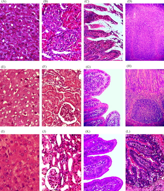
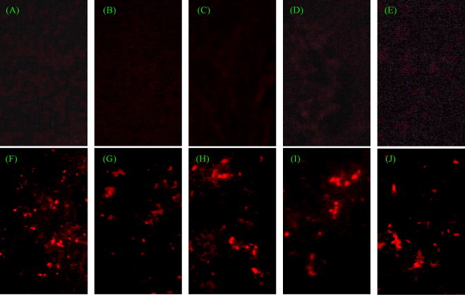
Transmissible gastroenteritis virus (TGEV) is the causative agent of porcine transmissible gastroenteritis (TGE), characterized by high mortality and severely retarded growth in piglets that dramatically affects the porcine industry. Previously, we have identified two shRNA-expressing plasmids pEGFP-U6/P1 and pEGFP-U6/P2 that target RNA-dependent RNA polymerase (RdRP) gene of TGEV with more than 95% of virus inhibition in vitro. In this study, inhibition of the TGEV replication by pEGFP-U6/P1 and pEGFP-U6/P2 was tested in mini-pigs. SPF mini-pigs at 25 days old were injected with the shRNA-expressing plasmids and then infected with TGEV. The results from the analyses of clinical signs, histopathology, indirect immunofluorescence (IIF) and RT-PCR show that the two shRNA-expressing plasmids could significantly decrease the quantity of TGEV in different organs and protect mini-pigs from TGEV infection. These findings illustrate the prospect for TGEV-specific shRNAs to be new anti-TGEV agents.
Crown Copyright 2010. Published by Elsevier B.V. All rights reserved.
Figures
Similar articles
-
Effective inhibition of porcine transmissible gastroenteritis virus replication in ST cells by shRNAs targeting RNA-dependent RNA polymerase gene.Antiviral Res. 2007 Apr;74(1):36-42. doi: 10.1016/j.antiviral.2006.12.007. Epub 2007 Jan 25. Antiviral Res. 2007. PMID: 17287033 Free PMC article.
-
In vitro inhibition of transmissible gastroenteritis coronavirus replication in swine testicular cells by short hairpin RNAs targeting the ORF 7 gene.Virol J. 2012 Aug 28;9:176. doi: 10.1186/1743-422X-9-176. Virol J. 2012. PMID: 22929207 Free PMC article.
-
(+)-Catechin inhibition of transmissible gastroenteritis coronavirus in swine testicular cells is involved its antioxidation.Res Vet Sci. 2015 Dec;103:28-33. doi: 10.1016/j.rvsc.2015.09.009. Epub 2015 Sep 12. Res Vet Sci. 2015. PMID: 26679792 Free PMC article.
-
An overview of immunological and genetic methods for detecting swine coronaviruses, transmissible gastroenteritis virus, and porcine respiratory coronavirus in tissues.Adv Exp Med Biol. 1997;412:37-46. doi: 10.1007/978-1-4899-1828-4_4. Adv Exp Med Biol. 1997. PMID: 9191988 Review.
-
Transmissible gastroenteritis virus infection: a vanishing specter.Dtsch Tierarztl Wochenschr. 2006 Apr;113(4):157-9. Dtsch Tierarztl Wochenschr. 2006. PMID: 16716052 Review.
Cited by
-
In vitro inhibition of porcine hemagglutinating encephalomyelitis virus replication with siRNAs targeting the spike glycoprotein and replicase polyprotein genes.Intervirology. 2012;55(1):53-61. doi: 10.1159/000323523. Epub 2011 Mar 3. Intervirology. 2012. PMID: 21372550 Free PMC article.
-
Inhibition of porcine transmissible gastroenteritis virus infection in porcine kidney cells using short hairpin RNAs targeting the membrane gene.Virus Genes. 2017 Apr;53(2):226-232. doi: 10.1007/s11262-016-1409-8. Epub 2016 Nov 15. Virus Genes. 2017. PMID: 27848068 Free PMC article.
-
Significant inhibition of re-emerged and emerging swine enteric coronavirus in vitro using the multiple shRNA expression vector.Antiviral Res. 2019 Jun;166:11-18. doi: 10.1016/j.antiviral.2019.03.010. Epub 2019 Mar 21. Antiviral Res. 2019. PMID: 30905822 Free PMC article.
-
Characterization and Evaluation of the Pathogenicity of a Natural Gene-Deleted Transmissible Gastroenteritis Virus in China.Transbound Emerg Dis. 2023 Mar 3;2023:2652850. doi: 10.1155/2023/2652850. eCollection 2023. Transbound Emerg Dis. 2023. PMID: 40303681 Free PMC article.
-
Plasmids Expressing shRNAs Specific to the Nucleocapsid Gene Inhibit the Replication of Porcine Deltacoronavirus In Vivo.Animals (Basel). 2021 Apr 23;11(5):1216. doi: 10.3390/ani11051216. Animals (Basel). 2021. PMID: 33922444 Free PMC article.
References
-
- Couzin J. RNAi safety comes under security. Science. 2006;312:1121. - PubMed
-
- de los Santos T., Wu Q., deAvila Botton S., Grubman M.J. Short hairpin RNA targeted to the highly conserved 2B nonstructural protein coding region inhibits replication of multiple serotypes of foot-and-mouth disease virus. Virology. 2005;335:222–231. - PubMed
MeSH terms
Substances
LinkOut - more resources
Full Text Sources
Research Materials
