. 2010 Apr 15;389(1-2):232-43.
doi: 10.1016/j.ijpharm.2010.01.019. Epub 2010 Jan 18.

Lipid-mediated delivery of RNA is more efficient than delivery of DNA in non-dividing cells

Affiliations

Lipid-mediated delivery of RNA is more efficient than delivery of DNA in non-dividing cells

S Zou et al. Int J Pharm. .

Abstract

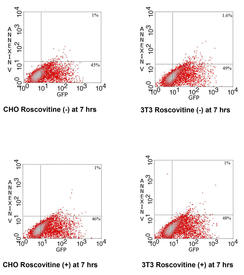
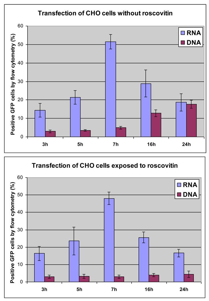
The design of appropriate gene delivery systems is essential for the successful application of gene therapy to clinical medicine. Cationic lipid-mediated delivery is a viable alternative to viral vector-mediated gene delivery in applications where transient gene expression is desirable. However, cationic lipid-mediated delivery of DNA to post-mitotic cells such as neurons is often reported to be of low efficiency, due to the presumed inability of the DNA to translocate to the nucleus. Lipid-mediated delivery of RNA is an attractive alternative to non-viral DNA delivery in some clinical applications, because transit across the nuclear membrane is not necessary. Here we report a comparative investigation of cationic lipid-mediated delivery of RNA versus DNA vectors encoding the reporter gene green fluorescent protein (GFP) in Chinese Hamster Ovary (CHO) and NIH3T3 cells following chemical inhibition of proliferation, and in primary mixed neuronal cell cultures. Using optimized formulations and transfection procedures, we assess gene expression by flow cytometry to specifically address some of the advantages and disadvantages of lipid-mediated RNA and DNA gene transfer. Despite inhibition of cell proliferation, over 45% of CHO cells express GFP after lipid-mediated transfection with RNA vectors. Transfection efficiency of DNA encoding GFP in proliferation-inhibited CHO cells was less than 5%. Detectable expression after RNA transfection occurs at least 3h earlier than after DNA transfection, but DNA transfection eventually produces a mean level of per cell GFP expression (as assayed by flow cytometry) that is higher than after RNA transfection. Transfection of proliferation-inhibited NIH3T3 cells and primary mixed neuronal cultures produced similar results, with RNA encoded GFP expression in 2-4 times the number of cells as after DNA encoded GFP expression. These results demonstrate the increased efficiency of RNA transfection relative to DNA transfection in non-dividing cells. We used firefly luciferase encoded by RNA and DNA vectors to investigate the time course of gene expression after delivery of RNA or DNA to primary neuronal cortical cells. Delivery of mRNA resulted in rapid onset (within 1h) of luciferase expression after transfection, a peak in expression 5-7h after transfection, and a return to baseline within 12h after transfection. After DNA delivery significant luciferase activity did not appear until 7h after transfection, but peak luciferase expression was always at least one order of magnitude higher than after RNA delivery. The peak expression after luciferase-expressing DNA delivery occurred 36-48 h after transfection and remained at a significant level for at least one week before dropping to baseline. This observation is consistent with our in vivo delivery results, which are shown as well. RNA delivery may therefore be more suitable for short-term transient gene expression due to rapid onset, shorter duration of expression and greater efficiency, particularly in non-dividing cells. Higher mean levels of expression per cell obtained following DNA delivery and the longer duration of expression confirm a continuing role for DNA gene delivery in clinical applications that require longer term transient gene expression.


Figures

Figure 1
Figure 1. FACScan analysis of cationic lipid-mediated RNA and DNA GFP transfection in CHO cells
Figure 1 is an example of the fraction of CHO cells transfected by RNA versus DNA in the absence of any inhibition of proliferation. Top panels show representative flow cytometry for RNA (left) versus DNA (right) transfections.
Figure 2
Figure 2. FACScan analysis of cationic lipid-mediated RNA GFP transfection of proliferation-inhibited CHO and NIH3T3 cells
Results are presented at the time point at which the maximum percentage of cells expresses GFP. Representative results show the percentage of GFP-expressing cells after RNA transfection at 7 hours in roscovitine treated (+) and untreated (−) CHO and NIH3T3 cells. In Figure 2, RNA Transfections, the two panels of flow cytometry results on the left hand side are CHO cells, while the right two panels are NIH3T3 cells. The CHO and NIH3T3 cells in the bottom two panels were treated with roscovitine to inhibit proliferation, while the top two panels show CHO and NIH3T3 cells in the absence of roscovitine. Each flow cytometry result shows Annexin V to detect early apoptosis on the vertical axis versus GFP on the horizontal axis. Figure 3 shows similar results for DNA GFP transfections in an identical presentation. Inhibition of proliferation has no effect on transfection with RNA on either cell type, but dramatically reduces the fraction of cells transfected with DNA vectors in both cell types in Figure 3.
Figure 3
Figure 3. FACScan analysis of cationic lipid-mediated DNA GFP transfection of proliferation-inhibited CHO and NIH3T3 cells
Representative results show the percentage of GFP-expressing CHO cells after DNA transfection 24 hours in roscovitine treated (+) and untreated (−) CHO and NIH3T3 cells.
Figure 4
Figure 4. Time course of GFP expression in CHO cells with and without proliferation-inhibition by roscovitine
Figure 4 and Figure 5 summarize the results of multiple transfections of GFP DNA versus RNA vectors over time. The percent of GFP positive cells by flow cytometry was plotted versus time in hours. Figure 4 shows the peak in fraction of CHO cells expressing GFP after transfection, and the absence of any effect from application of roscovitine using RNA, in contrast to the effect of roscovitine with DNA transfection.. Roscovitine has no effect on GFP RNA transfections, and the peak at 7 hours using RNA vectors is clearly illustrated in this Figure. DNA transfections, in contrast, reach only 18% in the absence of roscovitine, and less than 5% with proliferation-inhibition (roscovitine +). The fraction of cells which express GFP after GFP DNA transfection in the absence of treatment with roscovitine is still increasing after 24 hours, but is blocked at low level by roscovitine in the bottom panel.
Figure 5
Figure 5. DNA versus RNA GFP transfections in CHO and NIH3T3 cells with proliferation-inhibition
Figure 5 illustrates the effects of proliferation-inhibition with roscovitine in both CHO and NIH3T3 cells at 7 and 24 hours. By 24 hours the percent of CHO or NIH3T3 cells positive for GFP is already decreasing after RNA transfection, and is independent of roscovitine treatment. The fraction of cells expressing GFP after GFP DNA transfection in the absence of roscovitine is still increasing at 24 hours, but is blocked at a low level by roscovitine.
Figure 6
Figure 6. FACScan analysis of cationic lipid-mediated RNA and DNA transfection in primary mixed neuronal cells
Representative results show the percentage of GFP-expressing cells 7 hours and 24 hours post transfection, the time point of maximum percentage of cells expressing GFP, for RNA and DNA, respectively. Flow cytometry results (top panels) show that by 7 hours GFP RNA vectors transfected approximately 42% of primary neurons but only 0.3% GFP positive/Annexin negative (lower right quadrant) and 3.1% GFP positive/Annexin positive (upper right quadrant) after transfection with the GFP DNA vector in simultaneous transfections (top right panel).
Figure 7
Figure 7. Time course after RNA and DNA transfection in primary neuronal cells
Luciferase-encoding DNA and mRNA vectors were delivered to primary cortical neurons and analyzed. Delivery of mRNA resulted in a rapid onset of luciferase expression, a peak at 5–6 hours post-transfection, and return to base line by 12 hours after transfection. DNA gene expression peaked 36–48 hours after transfection, Averaged results of three wells at each time point for each experiment, with error bars, are given.
Figure 8
Figure 8. Representative examples luciferase immunohistochemistry after lipid-mediated RNA lipoplex gene delivery to rat brain via CSF injection
Figure 8 demonstrates the widespread distribution, uptake and expression that we have achieved after non-viral, cationic lipid-mediated gene delivery of mRNA vectors. Figures 8A and B demonstrate widespread subcortical expression at 40X magnification from two different animal experiments, with and without counterstaining, in which cells that are phenotypically neurons are clearly visible. Figure 8C is an example from another animal, at 20X magnification, and shows similar, widespread subcortical expression. Figure 8D is from an area of 8C at higher magnification. Figure 8E is another series from the same animal, and 8F is from an area of the same section at a 40X magnification. Figures 8G and 8H are control sections at 20 and 40X respectively.

Similar articles

Cited by

References

    1. Journal of Gene Medicine. 2008. Web site. http://www.wiley.co.uk/genetherapy/
    1. Allen MS, Wilgus JR, Chewning CS, Sayler GS, Simpson ML. A destabilized bacterial luciferase for dynamic gene expression studies. Syst Synth Biol. 2006;1:3–9. - PMC - PubMed
    1. Anderson DM, Hall LL, Ayyalapu A, Irion VR, Nantz MH, Hecker JG. Stability of mRNA/cationic lipid lipoplexes in human and rat cerebrospinal fluid: methods and evidence for non-viral mRNA gene delivery to the CNS. Human Gene Therapy. 2003;14:191–202. - PubMed
    1. Aronsohn AI, Hughes JA. Nuclear localization signal peptides enhance cationic liposome-mediated gene therapy. Journal of Drug Targeting. 1998;5:163–169. - PubMed
    1. Balasubramaniam RP, Bennett MJ, Aberle AM, Malone JG, Nantz MH, Malone RW. Structural and functional analysis of cationic transfection lipids: The hydrophobic domain. Gene Therapy. 1996;3:163–172. - PubMed

Publication types