. 2010 Apr;31(10):2816-26.
doi: 10.1016/j.biomaterials.2009.12.053. Epub 2010 Jan 18.

The effect of FGF-1 loaded alginate microbeads on neovascularization and adipogenesis in a vascular pedicle model of adipose tissue engineering

Affiliations

The effect of FGF-1 loaded alginate microbeads on neovascularization and adipogenesis in a vascular pedicle model of adipose tissue engineering

Monica L Moya et al. Biomaterials. 2010 Apr.

Abstract

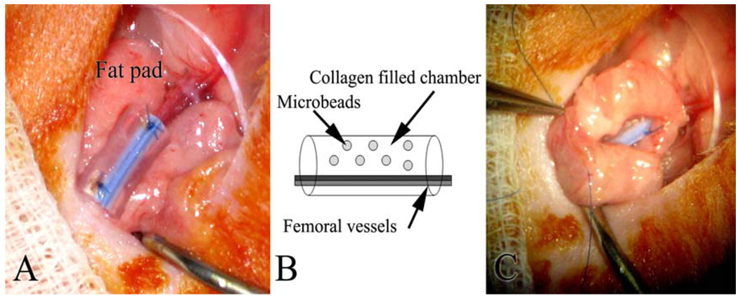
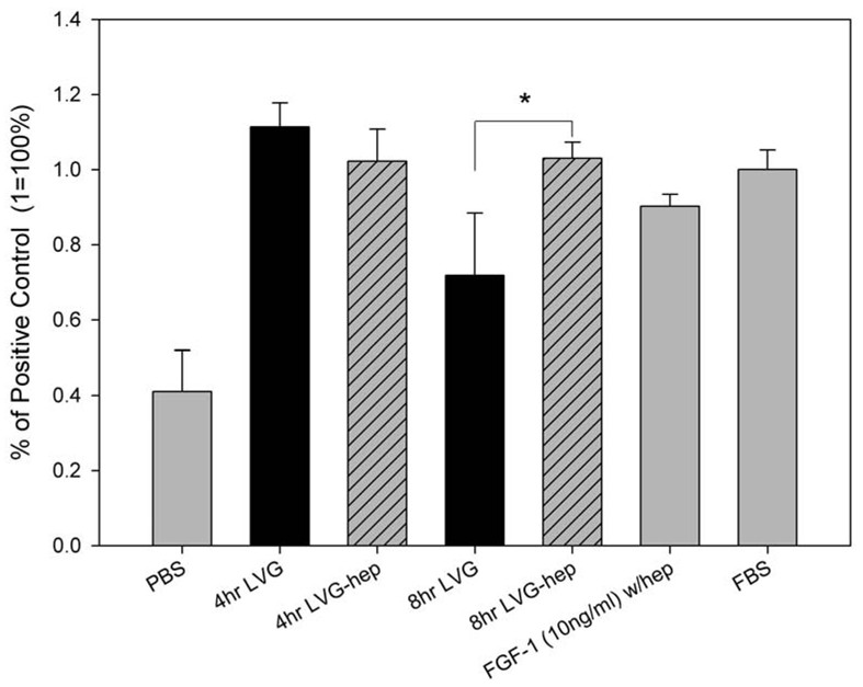
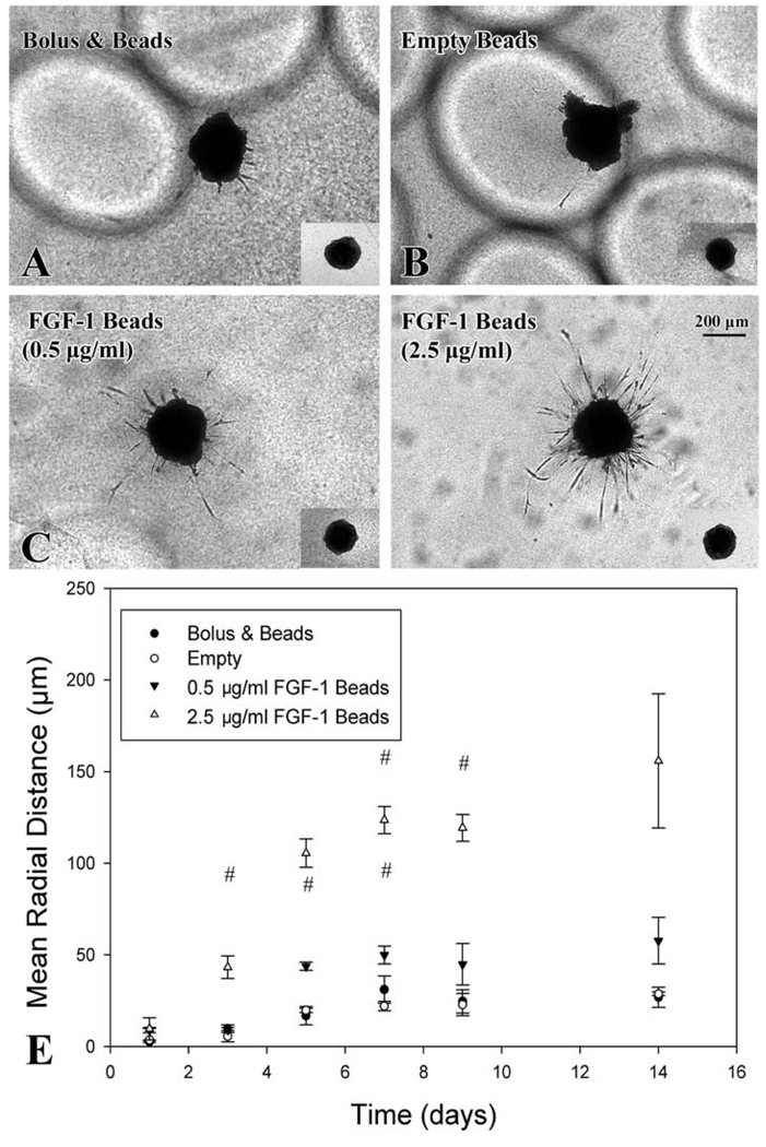
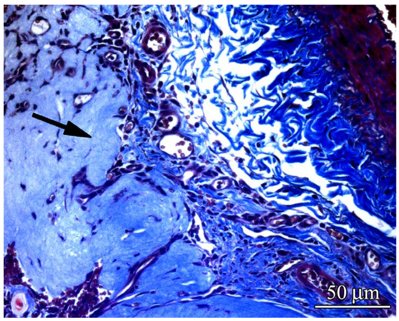
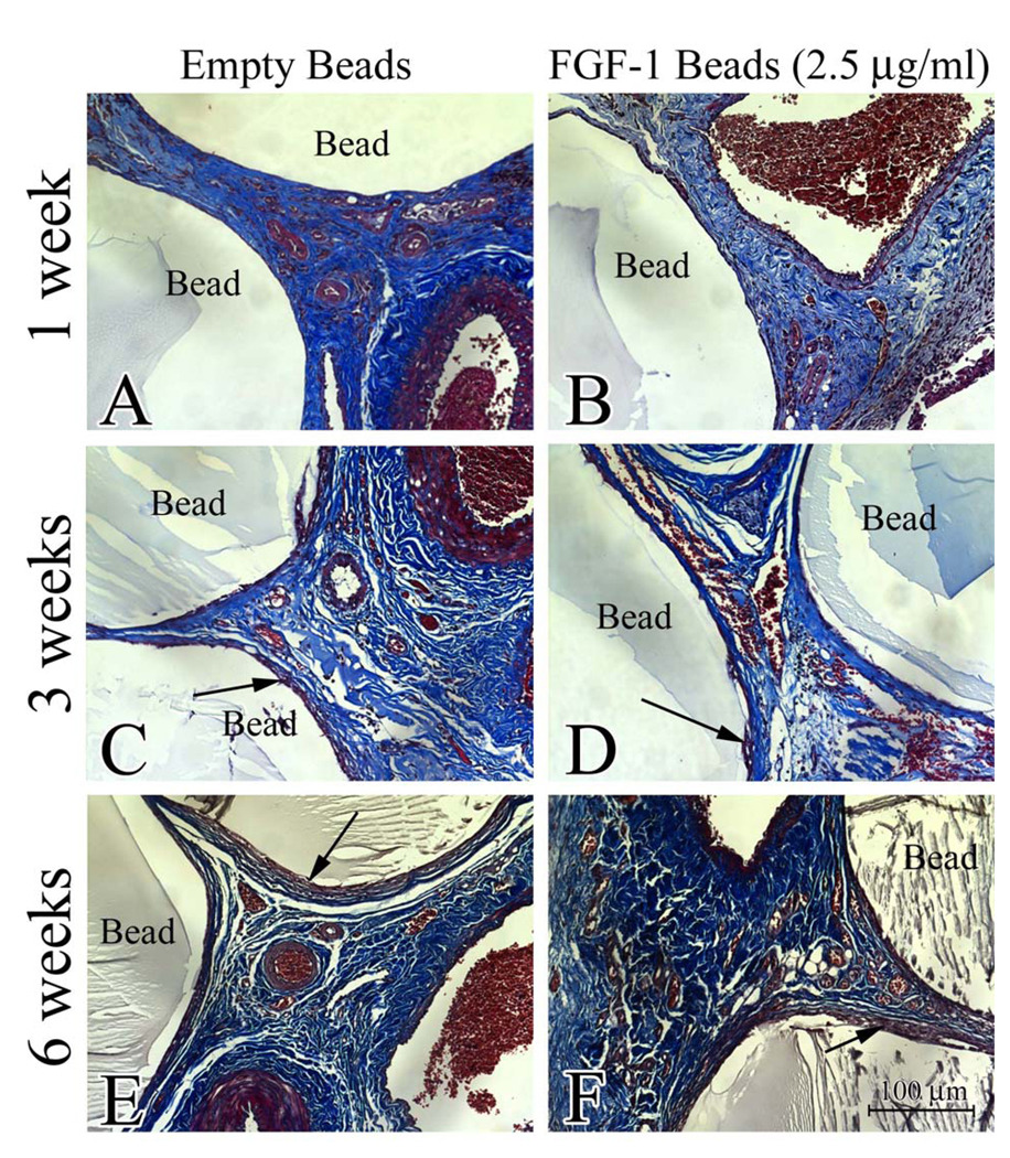
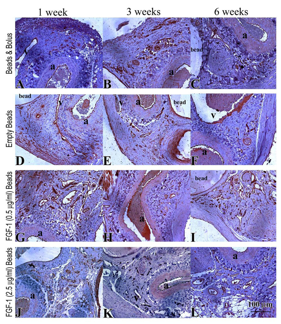
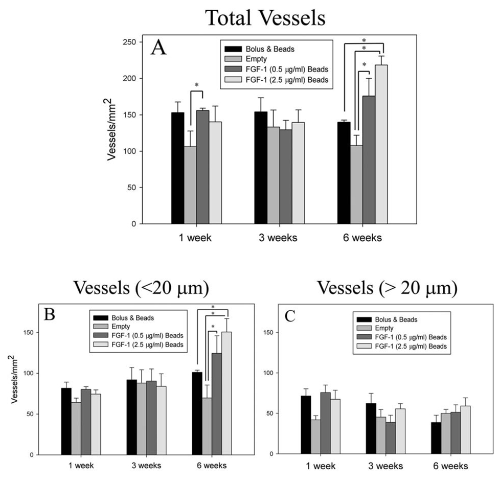
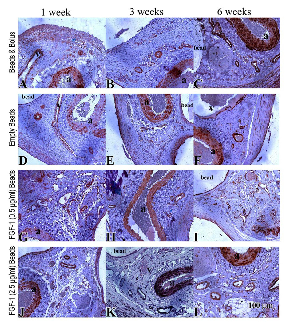
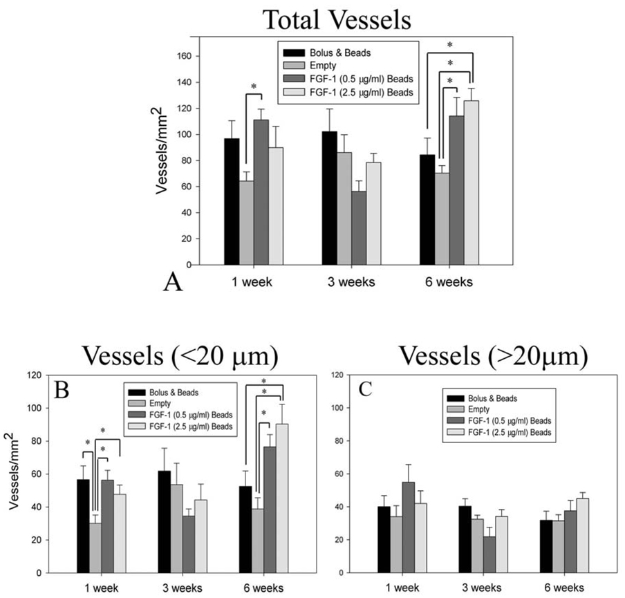
Engineered vascularized adipose tissue could serve as an alternative to traditional tissue reconstruction procedures. Adipose formation occurs in a coordinated fashion with neovascularization. Previous studies have shown that extracellular matrix-based materials supplemented with factors that stimulate neovascularization promote adipogenesis in a number of animal models. The present study examines the ability of fibroblast growth factor (FGF-1) delivered from alginate microbeads to induce neovascularization and adipogenesis in type I collagen gels in a vascular pedicle model of adipose tissue engineering. FGF-1 loaded microbeads stimulated greater vascular network formation in an in vitro 3D co-culture model than a single bolus of FGF-1. In in vivo studies, FGF-1 loaded beads suspended in collagen and implanted in a chamber surrounding the exposed femoral pedicle of a rat resulted in a significant increase in vascular density at 1 and 6 weeks in comparison to bolus administration of FGF-1. Staining for smooth muscle actin showed that over 48% of vessels had associated mural cells. While an increase in neovascularization was achieved, there was less than 3% adipose under any condition. These results show that delivery of FGF-1 from alginate beads stimulated a more persistent neovascularization response than bolus FGF-1 both in vitro and in vivo. However, unlike previous studies, this increased neovascularization did not result in adipogenesis. Future studies need to provide a better understanding of the relationship between neovascularization and adipogenesis in order to design advanced tissue engineering therapies.


Figures

Figure 1
Figure 1
Model of vascularized adipose tissue formation. A silicone tube is cut open and wrapped around exposed femoral artery and vein (A). The tube is then filled with a collagen gel containing alginate microbeads and sutured closed. (B). A local fat pad is wrapped around the tube to seal the ends (C).
Figure 2
Figure 2
FGF-1 released from alginate microbeads at 4 and 8 hours had significantly stimulated greater proliferation than negative control cells (PBS) (p<0.05, n=4). Black bars are FGF-1 released from alginate in the absence of heparin. Hashed bars are FGF-1 release from microbeads containing heparin. At 8 hours FGF-1 released from alginate beads containing heparin caused a significantly greater proliferation (*) than FGF-1 released from beads without heparin.
Figure 3
Figure 3
Sprouting angiogenesis from co-culture cell pellet stimulated by FGF-1 released from alginate microbeads. (A, B, C, D) Images taken at day 9 (inset images are of pellets at day 0) show vascular networks formation for groups with FGF-1 loaded beads. FGF-1 (2.5 µg/ml) beads showed increase and persistent vessel formation from day 3 to day 14 (E) # signifies statistical difference (p<0.05) from empty group.
Figure 4
Figure 4
Cellular and vessel invasion into the collagen gels could be seen at week 1. Samples harvested at 1 week and stained with Masson’s showed that collagen gel had been degraded and replaced fibrovascular tissue. (arrow indicates a region of non-degraded gel).
Figure 5
Figure 5
Sections of specimens were stained with Masson’s trichrome and imaged (20×, 0.17µm/pixel) to evaluate the presence of fibrosis around the alginate microbeads. No fibrosis was apparent at 1 week (A & D). Mild fibrosis (arrows) was visible at 6 weeks (C & F) regardless of whether beads were empty or loaded with FGF-1
Figure 6
Figure 6
CD31 stain of endothelial cells (brown) in specimens from rats treated for 1, 3 and 6 weeks with bolus FGF-1 (A–C), empty beads (D–F), FGF-1 (0.5 µg/ml) beads (G–I) FGF-1 (2.5 µg/ml) beads (J–L). The femoral artery and vein are indicated by “a” and “v” respectively.
Figure 7
Figure 7
Animals treated with FGF-1 loaded beads showed a higher total vessel number density (A) at 6 weeks. When the total vessels were separated into vessels less than 20µm (B) and vessels greater than 20 µm (C), the increase in the number of vessels appeared to primarily result from an increase in number of vessels less than 20 µm in diameter. * Indicates statistical significance p<0.05
Figure 8
Figure 8
SMA stain (brown) in specimens from rats treated for 1, 3 and 6 weeks with bolus FGF-1 (A–C), empty beads (D–F), FGF-1 (0.5 µg/ml) beads (G–I) FGF-1 (2.5 µg/ml) beads (J–L). Artery and vein are indicated by “a” and “v” respectively.
Figure 9
Figure 9
Animals treated with FGF-1 loaded beads showed a higher total number of SMA positive vessels per area (A) at 6 weeks. When the total vessels were separated into vessels less than 20µm (B) and vessels greater than 20 µm (C), the increase in the number of SMA positive vessels appeared to primarily result from an increase in number of positive vessels less than 20 µm in diameter. * Indicates statistical significance p<0.05

References

    1. Patrick CW. Breast tissue engineering. Annu Rev Biomed Eng. 2004;6:109–130. - PubMed
    1. Vacanti JP, Langer R. Tissue engineering: the design and fabrication of living replacement devices for surgical reconstruction and transplantation. Lancet. 1999;354 Suppl 1:SI32–SI34. - PubMed
    1. Griffith LG, Naughton G. Tissue engineering--current challenges and expanding opportunities. Science. 2002;295(5557):1009–1014. - PubMed
    1. Morrison WA. Progress in tissue engineering of soft tissue and organs. Surgery. 2009;145(2):127–130. - PubMed
    1. Uriel S, Brey EM, Greisler HP. Sustained low levels of fibroblast growth factor-1 promote persistent microvascular network formation. Am J Surg. 2006;192(5):604–609. - PubMed

Publication types

MeSH terms