

Nodal staging
- PMID: 20080453
- PMCID: PMC2821588
- DOI: 10.1102/1470-7330.2009.0017
Nodal staging
Abstract
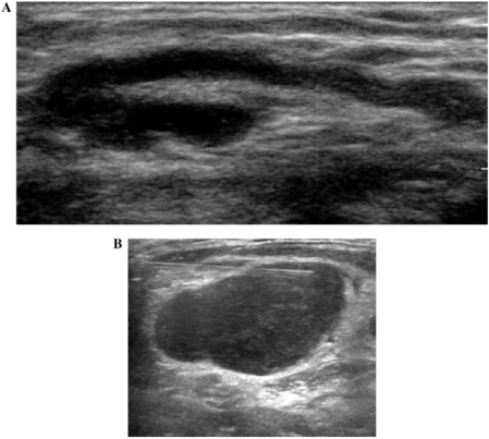
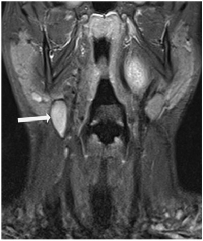
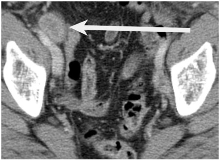
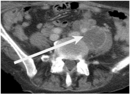
Lymph node metastases are a poor prognostic indicator in many tumours and therefore accurate identification during staging is important prior to commencing treatment. The presence of lymph node metastases can significantly alter patient management and therefore accurate diagnosis of the presence and extent of nodal disease can help optimise patient management. In this review, the radiologic features that aid in the differentiation of malignant and benign lymph nodes are discussed. The keys to successful interpretation on cross-sectional computed tomography (CT) and magnetic resonance imaging of nodal metastases are highlighted. The clinical role of positron emission tomography-CT imaging for nodal staging is discussed and emerging imaging techniques that may further improve nodal staging accuracy are surveyed.
Figures
References
-
- Steinkamp HJ, Cornehl M, Hosten N, Pegios W, Vogl T, Felix R. Cervical lymphadenopathy: ratio of long to short-axis diameter as a predictor of malignancy. Br J Radiol. 1995;68:266–70. doi:10.1259/0007-1285-68-807-266. PMid:7735765. - DOI - PubMed
-
- Na DG, Lim HK, Byun HS, Kim HD, Ko YH, Baek JH. Differential diagnosis of cervical lymphadenopathy: usefulness of color Doppler sonography. AJR Am J Roentgenol. 1997;168:1311–6. - PubMed
-
- Steinkamp HJ, Mueffelmann M, Böck JC, Thiel T, Kenzel P, Felix R. Differential diagnosis of lymph node lesions: a semiquantitataive approach with color Doppler ultrasound. Br J Radiol. 1998;71:828–33. - PubMed
-
- Choi MY, Lee JW, Jang KJ. Distinction between benign and malignant causes of cervical, axillary, and inguinal lymphadenopathy: value of Doppler spectral waveform analysis. AJR Am J Roentgenol. 1995;165:981–4. - PubMed
-
- Magarelli N, Guglielmi G, Savastano M, et al. Superficial inflammatory and primary neoplastic lymphadenopathy: diagnostic accuracy of power-Doppler sonography. Eur J Radiol. 2004;52:257–63. doi:10.1016/j.ejrad.2003.10.020. PMid:15544903. - DOI - PubMed
Publication types
MeSH terms
Substances
LinkOut - more resources
Full Text Sources
Other Literature Sources
Medical