. 2009 Dec 24;9(1):112-20.
doi: 10.1102/1470-7330.2009.0018.

Pathways of abdominal tumour spread: the role of the subperitoneal space

Affiliations

Pathways of abdominal tumour spread: the role of the subperitoneal space

Richard M Gore et al. Cancer Imaging. .

Abstract

The subperitoneal space is a large, unifying, anatomically continuous potential space that connects the peritoneal cavity with the retroperitoneum. This space is formed by the subserosal areolar tissue that lines the inner surfaces of the peritoneum and the musculature of the abdomen and pelvis. It contains the branches of the vascular, lymphatic, and nervous systems that supply the viscera. The subperitoneal space extends into the peritoneal cavity and is invested between the layers of the mesenteries and ligaments that support and interconnect the abdominal and pelvic organs. As such, it provides one large continuous space in which infectious, neoplastic, inflammatory, and hemorrhagic disease may spread in many directions.


Figures

Figure 1
Figure 1
Subperitoneal spaces. Frontal diagram of the posterior parietal wall of the upper abdomen shows the planes of peritoneal reflections that constitute the major ligaments and mesenteries of the subperitoneal space. Anatomic continuity between intraperitoneal structures and between extraperitoneal and intraperitoneal sites is established along the bare areas at the roots of origin of the supporting ligaments and mesenteries. FL, falciform ligament; RCL, right coronary ligament; BA, bare area of liver; HDL, hepatoduodenal ligament; LTL, left triangular ligament; GHL, gastrohepatic ligament; LS, lesser sac; GSL, gastrosplenic ligament; PL, phrenicocolic ligament; TM, transverse mesocolon; SBM, root of small bowel mesentery; DC, bare area of descending colon; AC, bare area of ascending colon. (From Meyers MA, Oliphant M, Berne AS, et al. The peritoneal ligaments and mesenteries: pathways of abdominal disease spread. Radiology 1987; 63: 594; with permission.)
Figure 2
Figure 2
Transverse mesocolon: anatomic relationships and planes of disease spread. (A) Frontal diagram shows the relationships of the transverse mesocolon (TM). The transverse mesocolon is continuous with the root of the small bowel mesentery (SBM), the splenorenal ligament (SRL), and the phrenicocolic ligament (PCL). (From Okino Y, Kiyosue H, Mori H, et al. Root of the small-bowel mesentery: correlative anatomy and CT features of pathologic conditions. Radiographics 2001; 21: 1480; with permission.) (B) Sagittal diagram through the transverse colon demonstrates preferential spread of pancreatic (P) disease through the transverse mesocolon, inferiorly along the taenia mesocolica–taenia libera (TL) haustra towards the TL–taenia omentalis (TO) row. This constitutes the inferior border of the transverse mesocolon. LS, lesser sac; S, stomach. (C) Fluid surrounds the middle colic vessels of the transverse mesocolon in this patient with pancreatitis. (From Meyers MA, Volberg R, Katzen B, et al. Haustral anatomy and pathology: a new look. II. Roentgen interpretation of pathologic alterations. Radiology 1973; 108: 505–12; with permission.)
Figure 3
Figure 3
Gastrocolic ligament: normal anatomy and pathology. (A) Sagittal diagram through the transverse colon demonstrates preferential spread of disease from the stomach (S), through the gastrocolic ligament, along the taenia omentalis (TO)–taenia mesocolica (TM) haustral row. This constitutes the superior border of the transverse colon. P, pancreas; TL, taenia libera. (From Meyers MA, Volberg R, Katzen B, et al. Haustral anatomy and pathology: a new look. II. Roentgen interpretation of pathologic alterations. Radiology 1973; 108: 505–12; with permission.) (B) Barium enema in a patient with Crohn disease demonstrates a fistula (arrow)from the transverse colon to the greater curvature aspect of the stomach via the gastrocolic ligament. (C) Direct invasion (arrows) of the superior aspect of the transverse colon along the gastrocolic ligament from a scirrhous carcinoma of the stomach.
Figure 4
Figure 4
Spread of gastric cancer into the gastrohepatic ligament. Coronal reformatted CT image shows tumour invasion of the gastrohepatic ligament (black arrow). Peritoneal tumour implants (white arrows) are identified as well.
Figure 5
Figure 5
Gastric cancer spread to the spleen via the gastrosplenic ligament. The subperitoneal space of the gastrosplenic ligament (arrows) is serving as a conduit of tumour spread in this patient with adenocarcinoma of the greater curvature of the stomach.
Figure 6
Figure 6
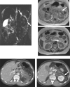
Inflammation of pancreatitis spreading from pancreas to left kidney via the splenorenal ligament: MR and CT findings in the same patient. (A) Coronal image from a magnetic resonance cholangiopancreatography examination shows high signal intensity fluid (arrow) in the perinephric space highlighting the lateral aspect of the left kidney. (B) Axial T1-weighted image shows low signal intensity between the pancreatic tail and left kidney (arrow). (C) Fat-suppressed T2-weighted axial image shows high signal intensity fluid lateral to the left kidney in the perinephric space. (D) CT scan shows low density necrosis in the pancreatic tail. (E) Scan obtained caudal to (D) shows fluid in the left perinephric space (arrow).
Figure 7
Figure 7
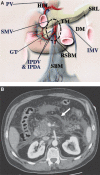
Small bowel mesentery: conduit of disease. (A) The anatomy near the root of the SBM (RSBM). The root of the SBM (area within dashed circle) is contiguous superiorly to the hepatoduodenal ligament (HDL) along the SMV, anteriorly to the transverse mesocolon (TM), and posterolaterally to the ascending mesocolon and descending mesocolon (DM). The gastrocolic trunk (GT) is a landmark of the junction between the transverse mesocolon and the root of the SBM. The inferior mesenteric vein (IMV) is a landmark of the descending mesocolon and joins the SMV or splenic vein on the left side of the root of the SBM. IPDA, inferior pancreaticoduodenal artery; IPDV, inferior pancreaticoduodenal vein; PV, portal vein; SRL, splenorenal ligament. (From Okino Y, Kiyosue H, Mori H, et al. Root of the small-bowel mesentery: correlative anatomy and CT features of pathologic conditions. Radiographics 2001; 21: 1476; with permission.) (B) CT scan shows fluid (arrow) in the subperitoneal space of the small bowel mesentery in this patient with pancreatitis.

Similar articles

Cited by

References

    1. Borley NR. In: Gray's anatomy. 40th. Philadelphia: WB Saunders; 2009. Peritoneum and peritoneal cavity; pp. 1347–1501.
    1. Oliphant M, Berne AS, Meyers MA. Subperitoneal spread of intra-abdominal disease. In: Meyers MA, editor. Computed tomography of the gastrointestinal tract. New York: Springer-Verlag; 1986. pp. 95–138.
    1. Oliphant M, Berne AS. Computed tomography of the subperitoneal space: demonstration of direct spread of intraabdominal disease. J Comput Assist Tomogr. 1982;6:1127–37. - PubMed
    1. Meyers MA, Oliphant M, Berne AS, et al. The peritoneal ligaments and mesenteries: pathways of intraabdominal spread of disease. Radiology. 1987;163:593–604. - PubMed
    1. Meyers MA. Intraperitoneal spread of infections. In: Meyers MA, editor. Dynamic radiology of the abdomen: normal and pathologic anatomy. 4th. New York: Springer-Verlag; 1993. pp. 55–114.

MeSH terms

LinkOut - more resources