. 2010 Jan 26;107(4):1630-5.
doi: 10.1073/pnas.0908953107. Epub 2009 Dec 28.

Alzheimer's-related endosome dysfunction in Down syndrome is Abeta-independent but requires APP and is reversed by BACE-1 inhibition

Affiliations

Alzheimer's-related endosome dysfunction in Down syndrome is Abeta-independent but requires APP and is reversed by BACE-1 inhibition

Ying Jiang et al. Proc Natl Acad Sci U S A. .

Abstract

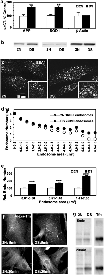
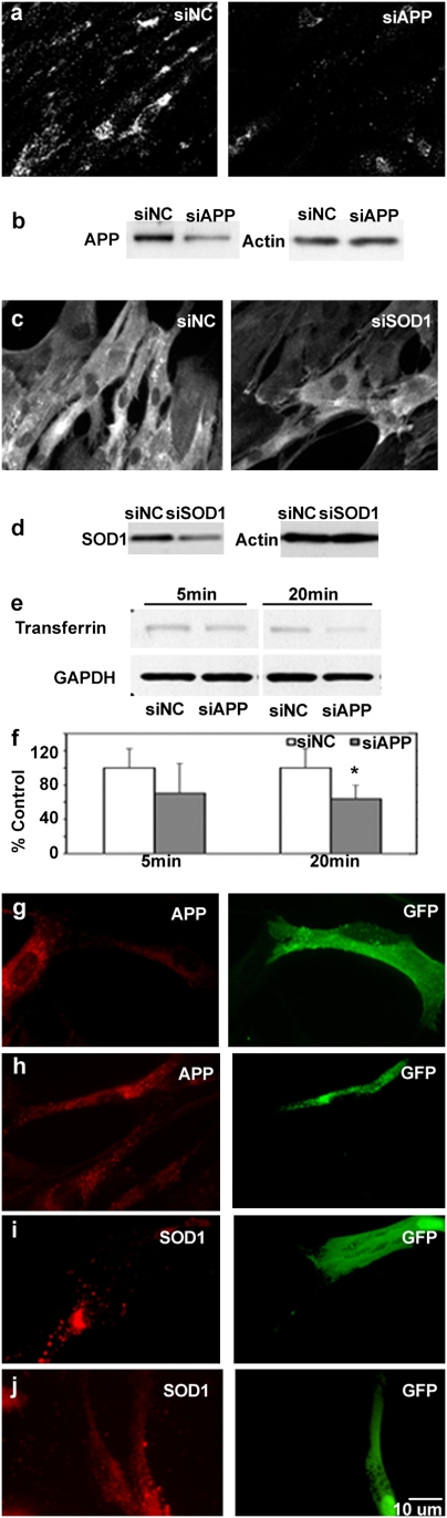
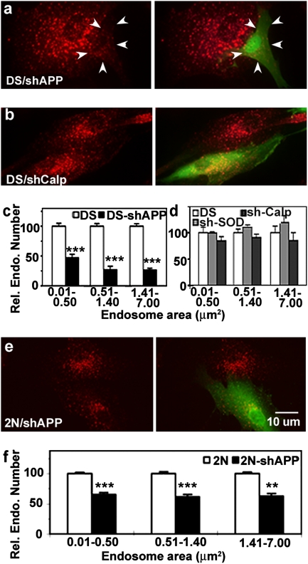
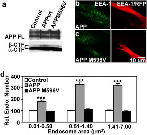
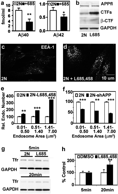
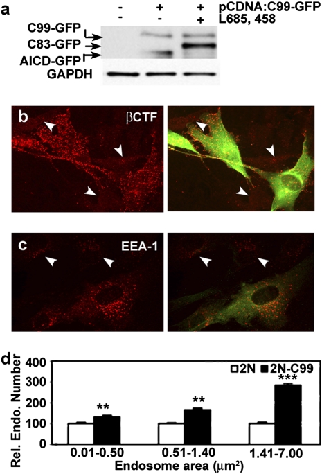
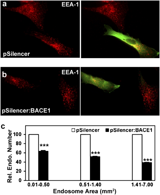
An additional copy of the beta-amyloid precursor protein (APP) gene causes early-onset Alzheimer's disease (AD) in trisomy 21 (DS). Endosome dysfunction develops very early in DS and AD and has been implicated in the mechanism of neurodegeneration. Here, we show that morphological and functional endocytic abnormalities in fibroblasts from individuals with DS are reversed by lowering the expression of APP or beta-APP-cleaving enzyme 1 (BACE-1) using short hairpin RNA constructs. By contrast, endosomal pathology can be induced in normal disomic (2N) fibroblasts by overexpressing APP or the C-terminal APP fragment generated by BACE-1 (betaCTF), all of which elevate the levels of betaCTFs. Expression of a mutant form of APP that cannot undergo beta-cleavage had no effect on endosomes. Pharmacological inhibition of APP gamma-secretase, which markedly reduced Abeta production but raised betaCTF levels, also induced AD-like endosome dysfunction in 2N fibroblasts and worsened this pathology in DS fibroblasts. These findings strongly implicate APP and the betaCTF of APP, and exclude Abeta and the alphaCTF, as the cause of endocytic pathway dysfunction in DS and AD, underscoring the potential multifaceted value of BACE-1 inhibition in AD therapeutics.


Conflict of interest statement

The authors declare no conflict of interest.

Figures

Fig. 1.
Fig. 1.
Assessment of DS fibroblasts (n = 5) compared with age-matched control samples (n = 5). (A) Expression levels of APP, SOD1, and α-actin in 2N and DS fibroblasts were determined by real-time qPCR (see Materials and Methods). **, P < 0.01. (B) Representative Western blot analysis of APP and SOD1 expression in DS and 2N fibroblasts. (C) Immunofluorescence images showing EEA1-labeled early endosomes in representative 2N and DS fibroblasts, with high magnification image shown in Inset. (D and E) The sizes of EEA1-positive vesicles in DS and 2N fibroblasts were counted with Image J, calculated, and graphed. Shown are the total number of EEA1-positive endosomes from 4 DS and 4 2N fibroblast lines and their distributions according to size (D) and the relative numbers of endosomes of different size ranges (E; mean ± SEM, n = 84; ***, P < 0.001.). Immunolabeling (F) and Western blot analysis (G) showing transferrin uptake by 2N and DS fibroblast at 5 and 20 min.
Fig. 2.
Fig. 2.
Down-regulation of APP and SOD1 expression in DS fibroblasts by siRNA and shRNA leads to reversal of endocytic dysfunction. (A) C1/6.1 immunolabeling shows DS fibroblasts transfected with siNC and siAPP. (B) Western blot analysis shows expression of APP in siAPP-treated DS fibroblasts. (C) Immunolabeling and (D) Western blot analysis with anti-SOD1 shows expression of SOD1 in siSOD1-transfected DS fibroblasts. (E) Representative Western blot analysis of transferrin uptake at 5 and 20 min in siAPP-treated DS fibroblasts. (F) Mean level of transferrin uptake from 3 experiments. *, P < 0.05. Expression of APP is shown in DS fibroblasts treated with shAPP (G) and vector (H). Expression of SOD1 is shown in shSOD1- (I) and vector- (J) transfected DS fibroblasts. In J the brightness levels of the red channel have been adjusted.
Fig. 3.
Fig. 3.
Altered early endosomal morphology in DS fibroblasts transfected with shAPP. Anti-GFP antibody (green) identifies transfected fibroblasts (arrowhead), whereas anti-EEA1 antibody (red) identifies early endosomes. Shown is the immunolabeling of DS fibroblasts transfected with shAPP (A) and shCalpastatin (B) with EEA1 (Left) and EEA1 plus GFP (Right). EEA1-positive endosomes in DS fibroblasts transfected with either shAPP (C) or shSOD1/shCalpastatin (D) were counted and analyzed with Image J. The bar graphs present the mean ± SEM for relative numbers of endosomes in different size ranges. n = 80, 55, and 50 for shAPP-, shSOD1-, and shCalpastatin-transfected DS fibroblasts, respectively. ***, P < 0.001. 2N fibroblasts also were treated with shAPP (E), and endosomal size was determined as above (F). n = 63; **, P < 0.01; *** P < 0.001.
Fig. 4.
Fig. 4.
APP overexpression in 2N fibroblasts induces AD-like endosome pathology. (A) Western blot of 2N fibroblast lysates collected 48 h after transfection with either dsRed-APP or dsRed-APPm596v was probed with antibody C1/6.1. Double-labeling for RFP and EEA1 shows EEA1-positive endosomes in APP- (B) and APPm596v- (C) transfected (RFP-positive) and nontransfected (RFP-negative) 2N fibroblasts. (D) Relative number of EEA1-positive endosomes of different size ranges in 2N fibroblasts overexpressing APP or APPm596v (mean ± SEM, n = 100). ***, P < 0.001.
Fig. 5.
Fig. 5.
γ-Secretase inhibition using L685,458 induces the AD-like endosomal phenotype in 2N fibroblasts. (A) Aβ-40 and Aβ-42 levels in cell-culture media collected 18 h after incubation with 10 μM of L685,458 were determined by ELISA (*, P < 0.05; **P < 0.01) . (B) Western blot analysis of cell lysates probed by C1/6.1 for APP and CTFs and by 6E10 for βCTF. Immunolabeling showed EEA1-positive vesicles in (C) DMSO- and (D) L685,458-treated fibroblasts. (E) The relative number of endosomes of different size ranges (mean ± SEM, n = 90; **, P < 0.01; ***, P < 0.001) in DMSO- and L685,458-treated 2N fibroblasts. (F) The relative number of endosomes of different size ranges (mean ± SEM, n = 74; **, P < 0.01; ***, P < 0.001) in shAPP-transfected and untransfected 2N fibroblasts. Western blot analysis (G) and its quantitation (H) of transferrin uptake in L685,458-treated versus control 2N fibroblasts normalized to GAPDH at 5 and 20 min. **, P < 0.01.
Fig. 6.
Fig. 6.
Overexpression of a βCTF construct induces the AD-like endosomal phenotype in 2N fibroblasts. (A) Western blot of C1/6.1of 2N fibroblasts transfected with pcDNA3:C99-GFP. Immunolabeling with anti-EEA1, N25, and anti-GFP antibodies shows βCTF (B) and EEA1 (C) in transfected and nontransfected (arrowhead) 2N fibroblasts. (D) Relative numbers of endosomes in transfected and nontransfected 2N fibroblasts (mean ± SEM, n = 55). **, P < 0.01; ***, P < 0.001.
Fig. 7.
Fig. 7.
Altered early endosomal morphology in DS fibroblasts transfected with pSilencer: BACE-1. Immunolabeling showed EEA1-positive vesicle in pSilencer- (A) and shBACE1- (B) transfected DS fibroblasts, with cotransfection of pGFP as a marker for transfection. The contrast levels have been adjusted in B. (C) Sizes of EEA1-positive vesicles in vector and shBACE1-transfected fibroblasts were calculated and graphed (mean ± SEM, n = 52). ***, P < 0.001.

Comment in

Similar articles

Cited by

References

    1. Haass C, et al. Amyloid beta-peptide is produced by cultured cells during normal metabolism. Nature. 1992;359:322–325. - PubMed
    1. Golde TE, Estus S, Younkin LH, Selkoe DJ, Younkin SG. Processing of the amyloid protein precursor to potentially amyloidogenic derivatives. Science. 1992;255:728–730. - PubMed
    1. Sisodia SS, Price DL. Role of the beta-amyloid protein in Alzheimer’s disease. FASEB J. 1995;9:366–370. - PubMed
    1. Athan ES, Lee JH, Arriaga A, Mayeux RP, Tycko B. Polymorphisms in the promoter of the human APP gene: Functional evaluation and allele frequencies in Alzheimer disease. Arch Neurol. 2002;59:1793–1799. - PubMed
    1. Rovelet-Lecrux A, et al. APP locus duplication causes autosomal dominant early-onset Alzheimer disease with cerebral amyloid angiopathy. Nat Genet. 2006;38:24–26. - PubMed

Publication types

MeSH terms