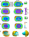
Model of human low-density lipoprotein and bound receptor based on cryoEM
- PMID: 20080547
- PMCID: PMC2798884
- DOI: 10.1073/pnas.0908004107
Model of human low-density lipoprotein and bound receptor based on cryoEM
Abstract
Human plasma low-density lipoproteins (LDL), a risk factor for cardiovascular disease, transfer cholesterol from plasma to liver cells via the LDL receptor (LDLr). Here, we report the structures of LDL and its complex with the LDL receptor extracellular domain (LDL.LDLr) at extracellular pH determined by cryoEM. Difference imaging between LDL.LDLr and LDL localizes the site of LDLr bound to its ligand. The structural features revealed from the cryoEM map lead to a juxtaposed stacking model of cholesteryl esters (CEs). High density in the outer shell identifies protein-rich regions that can be accounted for by a single apolipoprotein (apo B-100, 500 kDa) leading to a model for the distribution of its alpha-helix and beta-sheet rich domains across the surface. The structural relationship between the apo B-100 and CEs appears to dictate the structural stability and function of normal LDL.
Conflict of interest statement
The authors declare no conflict of interest.
Figures


Similar articles
-
Structure of apolipoprotein B100 bound to the low-density lipoprotein receptor.Nature. 2025 Feb;638(8051):829-835. doi: 10.1038/s41586-024-08223-0. Epub 2024 Dec 11. Nature. 2025. PMID: 39663455
-
Uptake of canine beta-very low density lipoproteins by mouse peritoneal macrophages is mediated by a low density lipoprotein receptor.J Biol Chem. 1986 Aug 25;261(24):11194-201. J Biol Chem. 1986. PMID: 3733751
-
Structure of triglyceride-rich human low-density lipoproteins according to cryoelectron microscopy.Biochemistry. 2003 Dec 23;42(50):14988-93. doi: 10.1021/bi0354738. Biochemistry. 2003. PMID: 14674775
-
Structure and physiologic function of the low-density lipoprotein receptor.Annu Rev Biochem. 2005;74:535-62. doi: 10.1146/annurev.biochem.74.082803.133354. Annu Rev Biochem. 2005. PMID: 15952897 Review.
-
Structure of apolipoprotein B-100 in low density lipoproteins.J Lipid Res. 2001 Sep;42(9):1346-67. J Lipid Res. 2001. PMID: 11518754 Review.
Cited by
-
Human low density lipoprotein: the mystery of core lipid packing.J Lipid Res. 2011 Feb;52(2):187-8. doi: 10.1194/jlr.E013417. Epub 2010 Dec 3. J Lipid Res. 2011. PMID: 21131533 Free PMC article. No abstract available.
-
Exploring the complete mutational space of the LDL receptor LA5 domain using molecular dynamics: linking SNPs with disease phenotypes in familial hypercholesterolemia.Hum Mol Genet. 2016 Mar 15;25(6):1233-46. doi: 10.1093/hmg/ddw004. Epub 2016 Jan 10. Hum Mol Genet. 2016. PMID: 26755827 Free PMC article.
-
Molecular modeling of LDLR aids interpretation of genomic variants.J Mol Med (Berl). 2019 Apr;97(4):533-540. doi: 10.1007/s00109-019-01755-3. Epub 2019 Feb 18. J Mol Med (Berl). 2019. PMID: 30778614 Free PMC article.
-
Selective regulation of macrophage lipid metabolism via nanomaterials' surface chemistry.Nat Commun. 2024 Sep 27;15(1):8349. doi: 10.1038/s41467-024-52609-7. Nat Commun. 2024. PMID: 39333092 Free PMC article.
-
Optimized negative staining: a high-throughput protocol for examining small and asymmetric protein structure by electron microscopy.J Vis Exp. 2014 Aug 15;(90):e51087. doi: 10.3791/51087. J Vis Exp. 2014. PMID: 25145703 Free PMC article.
References
-
- Havel RJ, Goldstein JL, Brown MS. Lipoproteins and Lipid Transport. In: Rosenberg PBaL., editor. The Metabolic Control of Disease. Philadelphia: W.B. Saunders Company; 1980. p. 398.
-
- Gaubatz JW, et al. Dynamics of dense electronegative low density lipoproteins and their preferential association with lipoprotein phospholipase A(2) J Lipid Res. 2007;48(2):348–357. - PubMed
-
- Brown MS, Goldstein JL. A receptor-mediated pathway for cholesterol homeostasis. Science. 1986;232(4746):34–47. - PubMed
-
- Hobbs HH, Brown MS, Goldstein JL. Molecular genetics of the LDL receptor gene in familial hypercholesterolemia. Hum Mutat. 1992;1(6):445–466. - PubMed
-
- Deckelbaum RJ, Shipley GG, Small DM, Lees RS, George PK. Thermal transitions in human plasma low density lipoproteins. Science. 1975;190(4212):392–394. - PubMed
Publication types
MeSH terms
Substances
Grants and funding
LinkOut - more resources
Full Text Sources

