. 2010 Jan 19;107(3):1235-40.
doi: 10.1073/pnas.0914140107. Epub 2009 Dec 29.

Mechanism of active targeting in solid tumors with transferrin-containing gold nanoparticles

Affiliations

Mechanism of active targeting in solid tumors with transferrin-containing gold nanoparticles

Chung Hang J Choi et al. Proc Natl Acad Sci U S A. .

Abstract

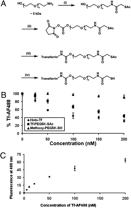
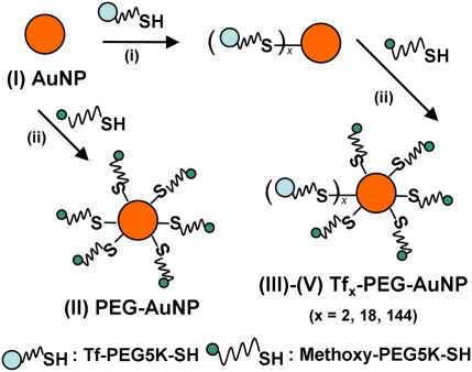
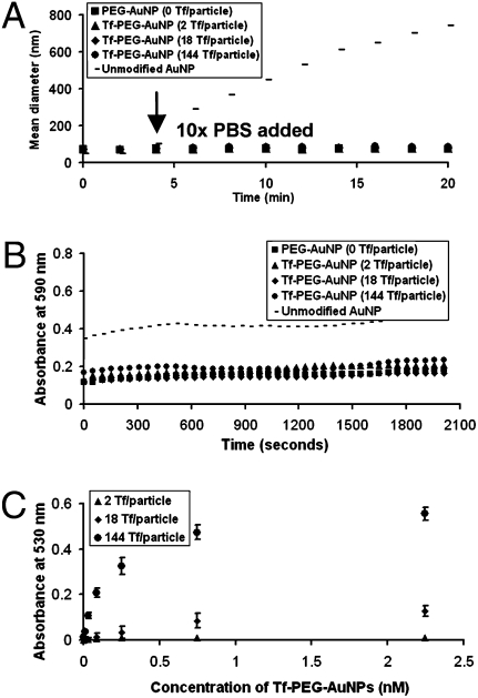
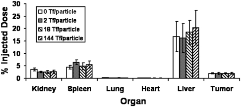
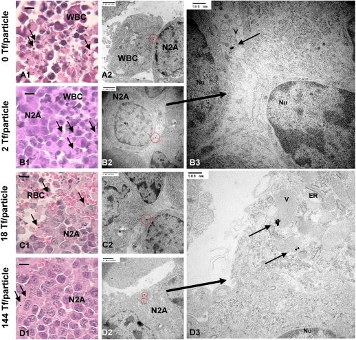
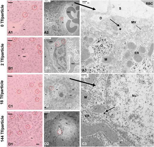
PEGylated gold nanoparticles are decorated with various amounts of human transferrin (Tf) to give a series of Tf-targeted particles with near-constant size and electrokinetic potential. The effects of Tf content on nanoparticle tumor targeting were investigated in mice bearing s.c. Neuro2A tumors. Quantitative biodistributions of the nanoparticles 24 h after i.v. tail-vein injections show that the nanoparticle accumulations in the tumors and other organs are independent of Tf. However, the nanoparticle localizations within a particular organ are influenced by the Tf content. In tumor tissue, the content of targeting ligands significantly influences the number of nanoparticles localized within the cancer cells. In liver tissue, high Tf content leads to small amounts of the nanoparticles residing in hepatocytes, whereas most nanoparticles remain in nonparenchymal cells. These results suggest that targeted nanoparticles can provide greater intracellular delivery of therapeutic agents to the cancer cells within solid tumors than their nontargeted analogs.


Conflict of interest statement

The authors declare no conflict of interest.

Figures

Fig. 1.
Fig. 1.
Synthesis and characterization of Tf-PEG-SAc. (A) Reaction conditions: (i) SATA, 4 h, DMF, room temperature; (ii) DSC, DMF/dioxane, 4 h, room temperature; (iii) Holo-Tf, 2 h, room temperature, 50 mM sodium phosphate (pH 7.4); (iv) hydroxylamine, 2 h, room temperature, 50 mM sodium phosphate, 25 mM EDTA (pH 7.5). Step iv renders free thiol groups for conjugation onto AuNPs to form Tf-PEG-AuNPs. (B) Competitive binding assay. PEGylation to form Tf-PEG-SAc did not drastically reduce the binding affinity of Tf. Reported data are expressed as the percent of fluorescence of cells incubated only with Tf-AF488. (C) Saturation binding of Tf-AF488 to Neuro2A cells. Error bars represent 1 SD from duplicate experiments.
Fig. 2.
Fig. 2.
Synthesis of Tf-PEG-AuNPs. Unmodified 50-nm AuNPs (I) were reacted with excess mPEG-SH to form PEG-AuNPs (II) as untargeted particles or first were reacted with various amounts of Tf-PEG-SH and later excess mPEG-SH to form Tf-PEG-AuNPs (III: 2 Tf per particle; IV: 18 Tf per particle; V: 144 Tf per particle).
Fig. 3.
Fig. 3.
Particle stability and binding. (A) In vitro stability against salt. Unmodified 50-nm AuNPs (I) showed instant aggregation after the addition of PBS at the fourth minute, but II–V remained stable. (B) In vitro stability against serum. PEGylation attenuated particle aggregation in 90% mouse serum. (C) Saturation binding of Tf-PEG-AuNPs to Neuro2A cells. Tf-PEG-AuNPs bound in a Tf content-dependent manner (III–V). Error bars indicate 1 SD from duplicate experiments.
Fig. 4.
Fig. 4.
In vivo organ distribution. Bulk particle localization in all organs was independent of Tf content. Gold contents are normalized to % injected dose. Error bars indicate 1 SD from each Tf-PEG-AuNP class (n = 3).
Fig. 5.
Fig. 5.
In vivo tumor tissue and intracellular distribution. (AD1) Light micrographs of “silver-enhanced” tumor sections. Arrows indicate “silver-enhanced” AuNPs. (Scale bar, 10 μm.) Independent of Tf content, most particles resided near leukocytes. Electron micrographs show particles either engulfed by leukocytes (A2) or tangentially touching Neuro2A cells (B–C2; enlarged image, B3). (D2 and enlarged image, D3) Particles with a high Tf content (V: 144 Tf per particle) can enter Neuro2A cells. ER, endoplasmic reticulum; M, mitochondrion; N2A, Neuro2A cell; Nu, nucleus; RBC, red blood cell; V, vesicle; WBC, leukocyte. [Scale bars: A1D2 (2 μm); B3 and D3 (500 nm).]
Fig. 6.
Fig. 6.
In vivo liver tissue and intracellular distribution. (AD1) Light micrographs of “silver-enhanced” liver sections. Arrows indicate “silver-enhanced” AuNPs. (Scale bar, 10 μm.) Independent of Tf content, particles rarely enter hepatocytes. Electron micrographs show particles either engulfed by Kupffer cells (A2) or residing in the space of Disse (A2; enlarged image, A3). Hepatocytes can internalize particles with high Tf contents (IV and V: CD2; enlarged image: C3). D, space of Disse; En, endothelial cell; ER, endoplasmic reticulum; G, Golgi apparatus; H, hepatocyte; HA, hepatic artery; KC and red circle, Kupffer cell; M, mitochondrion; MV, microvillus; Nu, nucleus; PV, hepatic portal vein; RBC, red-blood cell; S, sinusoid. [Scale bars: A1D2 (2 μm; A3 and C3 (500 nm).]

Similar articles

Cited by

References

    1. Davis ME, Chen ZG, Shin DM. Nanoparticle therapeutics: An emerging treatment modality for cancer. Nat Rev Drug Discov. 2008;7:771–782. - PubMed
    1. Matsumura Y, Maeda H. A new concept for macromolecular therapeutics in cancer chemotherapy: Mechanism of tumoritropic accumulation of proteins and the antitumor agent SMANCS. Cancer Res. 1986;46:6387–6392. - PubMed
    1. Dreher MR, et al. Tumor vascular permeability, accumulation, and penetration of macromolecular drug carriers. J Natl Cancer Inst. 2006;98:335–344. - PubMed
    1. Lopes de Menezes DE, Pilarski LM, Allen TM. In vitro and in vivo targeting of immunoliposomal doxorubicin to human B-cell lymphoma. Cancer Res. 1998;58:3320–3330. - PubMed
    1. Zhou Y, et al. Impact of single-chain Fv antibody fragment affinity on nanoparticle targeting of epidermal growth factor receptor-expressing tumor cells. J Mol Biol. 2007;371:934–947. - PMC - PubMed

Publication types

LinkOut - more resources