. 2010 Jan 19;107(3):1142-7.
doi: 10.1073/pnas.0910205107. Epub 2009 Dec 29.

Suppression of Alk8-mediated Bmp signaling cell-autonomously induces pancreatic beta-cells in zebrafish

Affiliations

Suppression of Alk8-mediated Bmp signaling cell-autonomously induces pancreatic beta-cells in zebrafish

Won-Suk Chung et al. Proc Natl Acad Sci U S A. .

Abstract

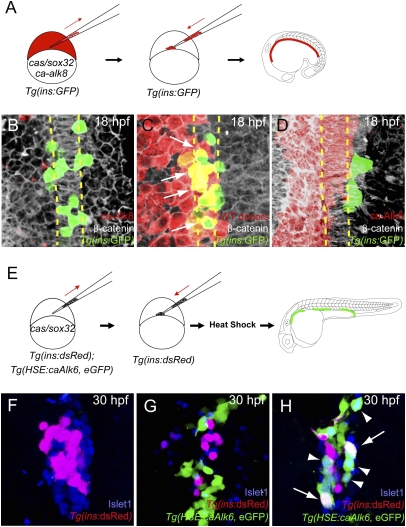
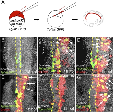
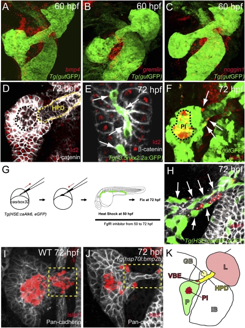
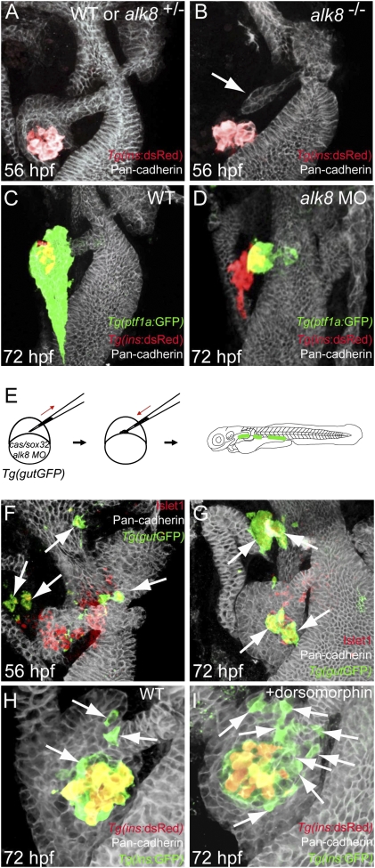
Bmp signaling has been shown to regulate early aspects of pancreas development, but its role in endocrine, and especially beta-cell, differentiation remains unclear. Taking advantage of the ability in zebrafish embryos to cell-autonomously modulate Bmp signaling in single cells, we examined how Bmp signaling regulates the ability of individual endodermal cells to differentiate into beta-cells. We find that specific temporal windows of Bmp signaling prevent beta-cell differentiation. Thus, future dorsal bud-derived beta-cells are sensitive to Bmp signaling specifically during gastrulation and early somitogenesis stages. In contrast, ventral pancreatic cells, which require an early Bmp signal to form, do not produce beta-cells when exposed to Bmp signaling at 50 hpf, a stage when the ventral bud-derived extrapancreatic duct is the main source of new endocrine cells. Importantly, inhibiting Bmp signaling within endodermal cells via genetic means increased the number of beta-cells, at early and late stages. Moreover, inhibition of Bmp signaling in the late stage embryo using dorsomorphin, a chemical inhibitor of Bmp receptors, significantly increased beta-cell neogenesis near the extrapancreatic duct, demonstrating the feasibility of pharmacological approaches to increase beta-cell numbers. Our in vivo single-cell analyses show that whereas Bmp signaling is necessary initially for formation of the ventral pancreas, differentiating endodermal cells need to be protected from exposure to Bmps during specific stages to permit beta-cell differentiation. These results provide important unique insight into the intercellular signaling environment necessary for in vivo and in vitro generation of beta-cells.


Conflict of interest statement

The authors declare no conflict of interest.

Figures

Fig. 1.
Fig. 1.
Activation of Bmp signaling cell-autonomously blocks the induction of dorsal bud-derived endocrine cells. (A) Schematic diagram of the cell transplantation protocol. Tg(ins:GFP) donors were injected with cas/sox32 and ca-alk8 RNA, along with rhodamine dextran, and the cells were transplanted into Tg(ins:GFP) hosts. (B–D) Ventral confocal images of Tg(ins:GFP) (green), β-catenin (white), and rhodamine dextran (red) at 18 hpf (the notochord is outlined by yellow dashed lines). (B and C) Tg(ins:GFP)-expressing cells are normally located close to the notochord in wild type (B) and hosts containing control donor cells (C). (D) Tg(ins:GFP) expression was absent in ca-Alk8-expressing endodermal cells. (E) Schematic diagram of the cell transplantation protocol. cas/sox32 overexpressing Tg(ins:dsRed); Tg(HSE:caAlk6, eGFP) donor cells were transplanted into Tg(ins:dsRed) hosts, and subsequently heat-shocked at various time points. (F–H) Confocal projection images of hosts with Tg(HSE:caAlk6, eGFP)-expressing cells (green) at 30 hpf after staining for dsRed (red) and Islet1 (blue), comparing control (F) and hosts [heat shock was applied at 9 (G) or 11 hpf (H)]. (G) When ca-alk6 expression was induced at 9 hpf, the number of Tg(ins:dsRed) and Islet1-expressing cells was reduced (compare F and G), and the Tg(HSE:caAlk6, eGFP)-expressing cells failed to express endocrine markers. (H) When ca-alk6 expression was induced at 11 hpf, several Tg(HSE:caAlk6, eGFP)-expressing cells expressed Islet1 (arrowheads) and some of them also coexpressed Tg(ins:dsRed) (arrows).
Fig. 2.
Fig. 2.
Suppression of Bmp signaling is sufficient to induce ectopic dorsal bud-derived pancreatic β-cells cell-autonomously. (A) Schematic diagram of the cell transplantation protocol. Tg(ins:GFP) donors were injected with cas/sox32 and dn-alk8 RNA along with rhodamine dextran (red), and the cells were transplanted into Tg(ins:GFP) hosts. (B–E) Ventral confocal images of Tg(ins:GFP) (green), β-catenin (white), and rhodamine dextran (red) at 18 hpf (the notochord is outlined by yellow dashed lines). (B) In control embryos, Tg(ins:GFP)-expressing cells are located close to the notochord. (C and D) Ectopic Tg(ins:GFP)-expressing cells (arrows) were found in lateral and anterior endodermal regions where dn-Alk8-expressing cells had incorporated, and all these ectopic cells were donor derived. (E) Gremlin1a overexpression in the endoderm also resulted in the ectopic formation of Tg(ins:GFP)-expressing cells (arrows). (F) When Hedgehog signaling was blocked with cyclopamine, Tg(ins:GFP)-expressing cells were almost absent in hosts containing control donor cells. (G) dn-Alk8 expression still induced the formation of Tg(ins:GFP)-expressing cells (arrows) even after cyclopamine treatment, and it did so cell-autonomously.
Fig. 3.
Fig. 3.
Activation of Bmp signaling cell-autonomously blocks the induction of ventral bud-derived endocrine cells. (A–C) Confocal images of Tg(gutGFP) (green) with mRNA expression (red) of bmp4 (A), gremlin1a (B), and noggin1 (C) at 60 hpf. (D and E) Confocal images of wild-type endoderm showing β-catenin (white) and Id2 (red) expression at 72 hpf. (D) Id2 expression is excluded from the hepatopancreatic duct cells (HPD; yellow dashed line) and appears to be downregulated or excluded from the pancreatic endocrine cells (PI, principal islet; black dashed line). (E) Id2 expression is also excluded from the intrapancreatic duct cells, labeled by Tg(-3.5nkx2.2a:GFP) expression (41) (green; arrows). (F) Confocal image of wild-type endoderm showing Tg(gutGFP) (green) and Islet1 (red) expression at 72 hpf. The ventral bud-derived endocrine cells (arrows) can be found at the junction between the pancreas and hepatopancreatic duct [the principal islet (PI) is outlined by black dashed line]. (G) Schematic diagram of the cell transplantation protocol. cas/sox32-overexpressing Tg(HSE:caAlk6, eGFP) donor cells were transplanted into wild-type hosts. Hosts were heat-shocked at 50 hpf and fixed at 72 hpf. After applying heat shock, hosts were treated with the Fgf receptor inhibitor SU5402, which induces ectopic Islet1-positive cells in the hepatopancreatic duct. (H) Single plane image of embryo stained for GFP (green), Pan-cadherin (white), and Islet1 (red) showing that Tg(HSE:caAlk6, eGFP)-expressing cells (arrows) fail to express Islet1 and display clear segregation from the ectopic Islet1-expressing cells (asterisks). (I and J) Effect of cell-nonautonomous activation of Bmp signaling by heat shock of Tg(hsp70l:bmp2b) at 50 hpf and subsequent treatment with SU5402 until fixation. Confocal projections of embryos stained for Pan-cadherin (white) and Islet1 (red) comparing control (I) and experimental (J) embryos. (I) Upon SU5402 treatment, ectopic Islet1-positive endocrine cells appeared in the hepatopancreatic duct (yellow dashed area), an effect that can be partially blocked by overexpression of Bmp2b (J). (K) Diagram of the endodermal organs at 72 hpf showing the liver (L; light red), pancreas (P; green), hepatopancreatic duct (HPD; yellow), gall bladder (GB; light yellow), and intestinal bulb (IB), as well as the ventral bud-derived endocrine cells (VBE; red) and principal islet (PI; red).
Fig. 4.
Fig. 4.
Suppression of Bmp signaling in a cell-autonomous fashion leads to an increase in ventral bud-derived endocrine cells. (A and B) Confocal projection of control (A) and alk8 mutant (B) endoderm at 56 hpf. Tg(ins:dsRed) embryos were stained for Pan-cadherin (white). (B) alk8 mutants exhibit a very small ventral pancreatic bud (arrow). (C and D) Confocal projections of control (C) and alk8 morphant (D) Tg(ptf1a:GFP);Tg(ins:dsRed) embryos stained for Pan-cadherin (white). In alk8 morphants (D), the ventral pancreas buds out but fails to expand and engulf the principal islet. (E) Schematic diagram of the cell transplantation protocol. Tg(gutGFP) donors were injected with cas/sox32 mRNA and alk8 splice-blocking MO, and the donor cells were transplanted into wild-type hosts. (F and G) Confocal projections of hosts containing alk8 morphant donor cells (green) stained for Pan-cadherin (white) and Islet1 (red) at 56 (F) and 72 (G) hpf. Most alk8 morphant donor cells gave rise to Islet1-expressing endocrine cells (arrows). (H and I) Confocal projections of Tg(ins:GFP);Tg(ins:dsRed) embryos stained for Pan-cadherin (white) comparing control (H) and dorsomorphin-treated (I) embryos. Compared with control embryos (H), dorsomorphin-treated embryos showed an increased number of GFP-only-positive β-cells (arrows) (I).

References

    1. Zaret KS, Grompe M. Generation and regeneration of cells of the liver and pancreas. Science. 2008;322:1490–1494. - PMC - PubMed
    1. Rossi JM, Dunn NR, Hogan BL, Zaret KS. Distinct mesodermal signals, including BMPs from the septum transversum mesenchyme, are required in combination for hepatogenesis from the endoderm. Genes Dev. 2001;15:1998–2009. - PMC - PubMed
    1. Wandzioch E, Zaret K. Dynamic signaling network for the specification of embryonic pancreas and liver progenitors. Science. 2009;324(5935):1707–1710. - PMC - PubMed
    1. Huang H, et al. Mypt1-mediated spatial positioning of Bmp2-producing cells is essential for liver organogenesis. Development. 2008;135(19):3209–3218. - PMC - PubMed
    1. Chung WS, Shin CH, Stainier DY. Bmp2 signaling regulates the hepatic versus pancreatic fate decision. Dev Cell. 2008;15:738–748. - PMC - PubMed

Publication types

MeSH terms

LinkOut - more resources