. 2010 Feb 23;107(8):3323-8.
doi: 10.1073/pnas.0905447107. Epub 2009 Dec 31.

Bioartificial matrices for therapeutic vascularization

Affiliations

Bioartificial matrices for therapeutic vascularization

Edward A Phelps et al. Proc Natl Acad Sci U S A. .

Abstract

Therapeutic vascularization remains a significant challenge in regenerative medicine applications. Whether the goal is to induce vascular growth in ischemic tissue or scale up tissue-engineered constructs, the ability to induce the growth of patent, stable vasculature is a critical obstacle. We engineered polyethylene glycol-based bioartificial hydrogel matrices presenting protease-degradable sites, cell-adhesion motifs, and growth factors to induce the growth of vasculature in vivo. Compared to injection of soluble VEGF, these matrices delivered sustained in vivo levels of VEGF over 2 weeks as the matrix degraded. When implanted subcutaneously in rats, degradable constructs containing VEGF and arginine-glycine-aspartic acid tripeptide induced a significant number of vessels to grow into the implant at 2 weeks with increasing vessel density at 4 weeks. The mechanism of enhanced vascularization is likely cell-demanded release of VEGF, as the hydrogels may degrade substantially within 2 weeks. In a mouse model of hind-limb ischemia, delivery of these matrices resulted in significantly increased rate of reperfusion. These results support the application of engineered bioartificial matrices to promote vascularization for directed regenerative therapies.


Conflict of interest statement

The authors declare no conflict of interest.

Figures

Fig. 1.
Fig. 1.
Design of PEGDA-based bioartificial matrices. (A) Bioactive ligands are functionalized with PEG-acrylate group: MMP-degradable cross-linker is functionalized with two PEG-acrylates, adhesive ligands and growth factors are mono-PEG-acrylated. Additon of a photoinitiator to an aqueous solution of PEGylated precursors results in formation of a cross-linked hydrogel after exposure to UV light by polymerization of acrylate end groups. (B) Biomolecules functionalized with PEG-acrylate have increased molecular weight when run on an SDS-PAGE gel. (i) Fluorescence of FITC label, Lane 1: RGD-FITC, Lane 2: A-PEG-RGD-FITC. (ii) BaCl/I stain for PEG, Lane 1: A-PEG-NHS, Lane 2 A-PEG-GPQ-PEG-A (iii) Coomassie, Lane 1: VEGF121-cys, Lane 2: A-PEG-VEGF121-cys. (C) Macroscopic view of a bioartificial PEGDA hydrogel cast in an 8 × 2 mm silicone mold.
Fig. 2.
Fig. 2.
In vitro results for degradation and cell spreading. (A) Degradation of bioartificial hydrogels incubated in collagenase-1 or MMP-2. Gels are seen to degrade after several hours of incubation, and degradation is decreased by addition of inhibitor and gels in PBS do not degrade. (B) Viability stain of NIH3T3 fibroblasts seeded in hydrogel formulations spread in bioartificial matrices with degradable sequences and adhesive sites: (i) type I collagen, (ii) PEGDA (iii) A-PEG-GPQ-PEG-A, and (iv) A-PEG-GPQ-PEG-A + A-PEG-RGD.
Fig. 3.
Fig. 3.
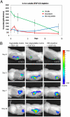
Degradation of subcutaneous implants containing ICG-labeled VEGF. (A) Quantification of VEGF fluorescent signal in implants shows early release for degradable matrix and late release for nondegradable matrix. Soluble injection shows continuous decline in signal strength. Initial soluble signal is significantly higher as it was not attenuated by subjection to cross-linking conditions. (B) Representative images from IVIS scanning of fluorescently labeled VEGF in degradable implants, nondegradable implants, and PBS injection. Number indicates average counts per unit area within ROI.
Fig. 4.
Fig. 4.
Micro-CT images of bioartificial matrices implanted subcutaneously in rats perfused with Microfil radio-opaque contrast agent. (A) GPQ + RGD + VEGF implants showing vasculature in surrounding tissue growing into implant, gray volume defines hydrogel. (B) Quantification of vascular volume/total implant volume. ± SEM (C) Representative scans from nondegradable implants with no adhesive ligands (PEGDA), nondegradable with adhesive ligands (PEGDA + RGD), degradable A-PEG-GPQ-PEG-A with no adhesive ligands (GPQ), degradable A-PEG-GPQ-PEG-A with adhesive A-PEG-RGD (GPQ + RGD), and degradable A-PEG-GPQ-PEG-A with adhesive A-PEG-RGD and A-PEG-VEGF (GPQ + RGD + VEGF) N = 5.
Fig. 5.
Fig. 5.
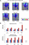
Hind-limb perfusion in mice with ligated femoral artery and vein. (A) LDPI imaging of limb perfusion at day 7 responding to treatment conditions: no treatment, PBS injection, soluble A-PEG-VEGF121-cys injection, degradable A-PEG-GPQ-PEG-A with adhesive A-PEG-RGDand degradable A-PEG-GPQ-PEG-A with adhesive A-PEG-RGD and A-PEG-VEGF121-cys. (B) Quantification of perfusion ratio (normal leg : ischemic leg) at days 4 and 7 postsurgery. Error bars represent standard error of the mean (N = 10).

References

    1. Peripheral Artery Disease Quick Facts. American Heart Association; 2009.
    1. Heart Disease and Stroke Statistics—2009 Update. American Heart Association; 2009.
    1. van Weel V, van Tongeren RB, van Hinsbergh VWM, van Bockel JH, Quax PHA. Vascular growth in ischemic limbs: A review of mechanisms and possible therapeutic stimulation. Ann Vasc Surg. 2008;22:582–597. - PubMed
    1. Hummers LK, Hall A, Wigley FM, Simons M. Evidence for abnormal angiogenesis in scleroderma patients. Arthritis and Rheum. 2004;50:S630–S631.
    1. Annex BH, Simons M. Growth factor-induced therapeutic angiogenesis in the heart: Protein therapy. Cardiovasc Res. 2005;65:649–655. - PubMed

Publication types

MeSH terms

LinkOut - more resources