. 2010 Jan 26;107(4):1425-30.
doi: 10.1073/pnas.0911996107. Epub 2010 Jan 5.

Establishment of a neuroepithelial barrier by Claudin5a is essential for zebrafish brain ventricular lumen expansion

Affiliations

Establishment of a neuroepithelial barrier by Claudin5a is essential for zebrafish brain ventricular lumen expansion

Jingjing Zhang et al. Proc Natl Acad Sci U S A. .

Abstract

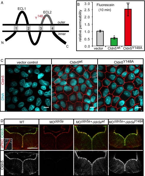
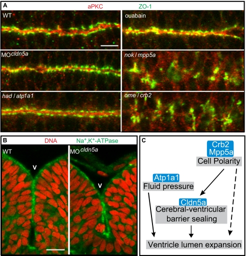
Lumen expansion driven by hydrostatic pressure occurs during many morphogenetic processes. Although it is well established that members of the Claudin family of transmembrane tight junction proteins determine paracellular tightness within epithelial/endothelial barrier systems, functional evidence for their role in the morphogenesis of lumenized organs has been scarce. Here, we identify Claudin5a as a core component of an early cerebral-ventricular barrier system that is required for ventricular lumen expansion in the zebrafish embryonic brain before the establishment of the embryonic blood-brain barrier. Loss of Claudin5a or expression of a tight junction-opening Claudin5a mutant reduces brain ventricular volume expansion without disrupting the polarized organization of the neuroepithelium. Perfusion experiments with the electron-dense small molecule lanthanum nitrate reveal that paracellular tightness of the cerebral-ventricular barrier decreases upon loss of Claudin5a. Genetic analyses show that the apical neuroepithelial localization of Claudin5a depends on epithelial cell polarity and provide evidence for concerted activities between Claudin5a and Na(+),K(+)-ATPase during luminal expansion of brain ventricles. These data establish an essential role of a barrier-forming Claudin in ventricular lumen expansion, thereby contributing to brain morphogenesis.


Conflict of interest statement

The authors declare no conflict of interest.

Figures

Fig. 1.
Fig. 1.
Loss of Claudin5a disrupts the neuroepithelial paracellular barrier function. (A and A′) Whole-mount in situ hybridization of claudin5a (cldn5a) expression at two developmental time points. Dorsal views onto the brain are shown magnified within the insets. (A) Before brain ventricle formation (14 hpf), cldn5a is strongly expressed within the developing central nervous system including the hindbrain (hb) and spinal cord (sc). Weaker expression of cldn5a is found within the dorsal midbrain region (mb). (A′) During ventricle expansion (30 hpf), cldn5a is strongly expressed within the spinal cord and the neuroepithelial ventricular zones of ventral hindbrain and midbrain. There is also strong expression in the forebrain (fb) ventricular zone. (B) Confocal microscopic images of cross-sections through the trunk region. Strong expression of Cldn5 proteins is present within the spinal cord (sc) and the dorsal aorta (da) but not the cardinal vein (cv) at 30 hpf. (C) Injection of MOcldn5a efficiently blocks expression of Cldn5a protein within the apical neuroepithelium lining the brain ventricles (V). Endothelial expression of Cldn5b is not affected in cldn5a morphants (arrows). (DF) Electron micrographs of neuroepithelial cells covering the cerebral ventricles (V) after intraventricular injection of the electron-dense molecule lanthanum nitrate. In the WT, paracellular clefts are tight for the tracer, which accumulates in a dot-like pattern at the TJ (arrows). Knock-down of cldn5a results in diffusion of lanthanum nitrate into the paracellular space between cells (arrows). In rescue embryos, in which the cldn5a knock-down was rescued by concomitant cldn5a mRNA injection, electron dense material is confined to apico-lateral membranes of neuroepithelial cells similar to the distribution in WT embryos. (Scale bars: B and C, 50 μm; D, 2 μm.)
Fig. 2.
Fig. 2.
The TJ opening Claudin5aY148A mutant increases TJ permeability. (A) Schematic representation of the four-transmembrane pass TJ protein Claudin5a (Cldn5a). The two extracellular loops (ECL), facing the paracellular space, are involved in claudin-claudin trans interactions. The position of an aromatic residue within ECL2, which is conserved between all classic claudins (17) and which is essential for trans interactions, is highlighted in red. ECL2, which is conserved between all classic claudins (17) and which is essential for trans interactions. (B) Stable transfection of murine Cldn5wt reduces the paracellular permeability of fluorescein (filter culture) compared to the vector control, whereas transfection of the mutant Cldn5Y148A causes an increase of fluorescein permeability. Data represent mean ± SEM, n ≥ 10; *, P < 0.05, **, P < 0.01. (C) Stable transfection of murine Cldn5wt and Cldn5Y148A into MDCK-II cells. The Cldn5 variants are strongly enriched at cell-cell contacts as detected by immunocytochemistry against the N-terminal FLAG-tag. (Scale bars: 10 μm.) (D) Injection of mRNA encoding zebrafish Cldn5awt or Cldn5aY148A into cldn5a morphants results in the correct localization of the proteins at the ventricular surface of the 30 hpf neural tube as detected by confocal microscopy of sectioned immunohistochemical stainings. (Scale bar: 50 μm.)
Fig. 3.
Fig. 3.
Loss of Claudin5a affects expansion of the brain ventricular lumen at 20 hpf. (AE) Shown are confocal microscopic Z-scans of in vivo sodium green indicator-labeled embryos. The ventricular lumen is indicated by false-coloring (orange). Indicated are ventricles of the midbrain (MBV) and hindbrain (HBV). (F) Quantifications of ventricular volume were generated from detailed 3D-reconstructions of confocal Z-scan sections for a total of 4–5 embryos per sample group by using Volocity software (unit size is [×106 μm3]). Data represent mean ± SEM, n ≥ 4, *, P = 0.016 in all three cases. (Scale bar: 100 μm.)
Fig. 4.
Fig. 4.
Loss of Claudin5a does not affect neuroepithelial tissue integrity. (A) Shown are apical views of immunohistochemical stainings onto the hindbrain ventricular zone of cell polarity mutants, Cldn5a- or Atp1a1-deficient embryos, or ouabain-treated embryos at 30 hpf. The neuroepithelial localization of Zonula occludens-1 (ZO-1) or aPKC is only affected in the cell polarity mutants nokm520/mpp5a and omem289/crb2. (Scale bar: 20 μm.) (B) Loss of Cldn5a does not affect the neuroepithelial localization of Na+,K+-ATPase which is labeled with the a6F antibody. Shown are confocal microscopic images of sections of immunohistochemical stainings of the hindbrain and ventricle. (Scale bar: 20 μm.) (C) Schematic diagram of developmental/cellular processes contributing to brain ventricle expansion. Our study suggests that the cell polarity regulators Crb2 and Mpp5a are essential for neuroepithelial integrity and maintenance of the TJ. Moreover, Crumbs complex proteins may be directly required for lumen formation (Results and Discussion). Tightness of the TJ is, at least in part, regulated by Cldn5a, which seals the neuroepithelial layer to maintain the fluid pressure, which may depend on the ion pump activity of Atp1a1. Ventricular fluid accumulation drives expansion of brain ventricles and tissue morphogenesis. crumbs2 (crb2); heart and mind (had); membrane protein, palmitoylated 5a (mpp5a); nagie oko (nok); oko meduzy (ome); Ventricle (V).

Similar articles

Cited by

References

    1. Lowery LA, Sive H. Initial formation of zebrafish brain ventricles occurs independently of circulation and requires the nagie oko and snakehead/atp1a1a.1 gene products. Development. 2005;132:2057–2067. - PubMed
    1. Jeong JY, et al. Functional and developmental analysis of the blood-brain barrier in zebrafish. Brain Res Bull. 2008;75:619–628. - PubMed
    1. Bill BR, et al. Development and Notch signaling requirements of the zebrafish choroid plexus. PLoS One. 2008;3:e3114. - PMC - PubMed
    1. García-Lecea M, Kondrychyn I, Fong SH, Ye ZR, Korzh V. In vivo analysis of choroid plexus morphogenesis in zebrafish. PLoS One. 2008;3:e3090. - PMC - PubMed
    1. Furuse M, Tsukita S. Claudins in occluding junctions of humans and flies. Trends Cell Biol. 2006;16:181–188. - PubMed

Publication types

MeSH terms