

Mammalian Mst1 and Mst2 kinases play essential roles in organ size control and tumor suppression
- PMID: 20080598
- PMCID: PMC2824397
- DOI: 10.1073/pnas.0911409107
Mammalian Mst1 and Mst2 kinases play essential roles in organ size control and tumor suppression
Abstract
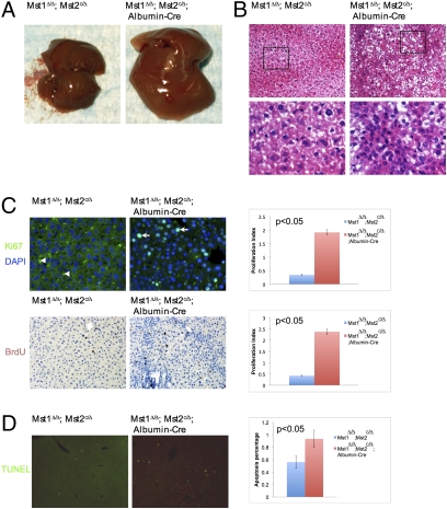
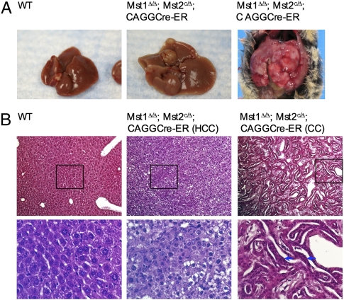
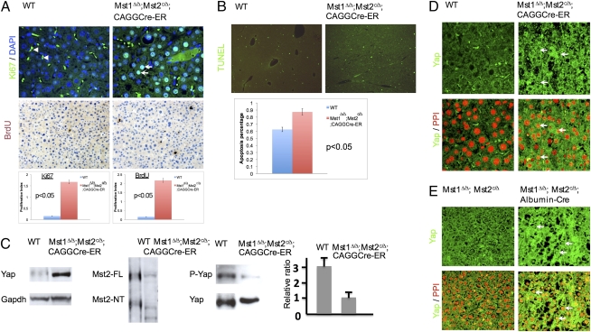
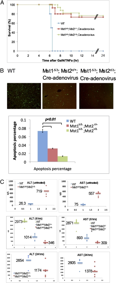
Control of organ size by cell proliferation and survival is a fundamental developmental process, and its deregulation leads to cancer. However, the molecular mechanism underlying organ size control remains elusive in vertebrates. In Drosophila, the Hippo (Hpo) signaling pathway controls organ size by both restricting cell growth and proliferation and promoting cell death. Here we investigated whether mammals also require the Hpo pathway to control organ size and adult tissue homeostasis. We found that Mst1 and Mst2, the two mouse homologs of the Drosophila Hpo, control the sizes of some, but not all organs, in mice, and Mst1 and Mst2 act as tumor suppressors by restricting cell proliferation and survival. We show that Mst1 and Mst2 play redundant roles, and removal of both resulted in early lethality in mouse embryos. Importantly, tumors developed in the liver with a substantial increase of the stem/progenitor cells by 6 months after removing Mst1 and Mst2 postnatally. We show that Mst1 and Mst2 were required in vivo to control Yap phosphorylation and activity. Interestingly, apoptosis induced by TNFalpha was blocked in the Mst1 and Mst2 double-mutant cells both in vivo and in vitro. As TNFalpha is a pleiotropic inflammatory cytokine affecting most organs by regulating cell proliferation and cell death, resistance to TNFalpha-induced cell death may also contribute significantly to tumor formation in the absence of Mst1 and Mst2.
Conflict of interest statement
The authors declare no conflict of interest.
Figures
Similar articles
-
Hippo signaling is a potent in vivo growth and tumor suppressor pathway in the mammalian liver.Proc Natl Acad Sci U S A. 2010 Jan 26;107(4):1437-42. doi: 10.1073/pnas.0911427107. Epub 2010 Jan 4. Proc Natl Acad Sci U S A. 2010. PMID: 20080689 Free PMC article.
-
MOBKL1A/MOBKL1B phosphorylation by MST1 and MST2 inhibits cell proliferation.Curr Biol. 2008 Mar 11;18(5):311-21. doi: 10.1016/j.cub.2008.02.006. Curr Biol. 2008. PMID: 18328708 Free PMC article.
-
Functional role of Mst1/Mst2 in embryonic stem cell differentiation.PLoS One. 2013 Nov 5;8(11):e79867. doi: 10.1371/journal.pone.0079867. eCollection 2013. PLoS One. 2013. PMID: 24224013 Free PMC article.
-
The functions of the Hippo signaling pathway in immune cells.Yi Chuan. 2017 Jul 20;39(7):650-658. doi: 10.16288/j.yczz.17-083. Yi Chuan. 2017. PMID: 28757479 Review.
-
The DeMSTification of mammalian Ste20 kinases.Curr Biol. 2009 May 26;19(10):R421-5. doi: 10.1016/j.cub.2009.04.022. Curr Biol. 2009. PMID: 19467213 Review.
Cited by
-
Integrin α2β1 inhibits MST1 kinase phosphorylation and activates Yes-associated protein oncogenic signaling in hepatocellular carcinoma.Oncotarget. 2016 Nov 22;7(47):77683-77695. doi: 10.18632/oncotarget.12760. Oncotarget. 2016. PMID: 27765911 Free PMC article.
-
Hippo signaling regulates differentiation and maintenance in the exocrine pancreas.Gastroenterology. 2013 Jun;144(7):1543-53, 1553.e1. doi: 10.1053/j.gastro.2013.02.037. Epub 2013 Feb 27. Gastroenterology. 2013. PMID: 23454691 Free PMC article.
-
Yap1 protein regulates vascular smooth muscle cell phenotypic switch by interaction with myocardin.J Biol Chem. 2012 Apr 27;287(18):14598-605. doi: 10.1074/jbc.M111.329268. Epub 2012 Mar 12. J Biol Chem. 2012. PMID: 22411986 Free PMC article.
-
The Hippo Signaling Pathway in Cardiac Development and Diseases.Front Cell Dev Biol. 2019 Oct 1;7:211. doi: 10.3389/fcell.2019.00211. eCollection 2019. Front Cell Dev Biol. 2019. PMID: 31632964 Free PMC article. Review.
-
YAP antagonizes TEAD-mediated AR signaling and prostate cancer growth.EMBO J. 2023 Feb 15;42(4):e112184. doi: 10.15252/embj.2022112184. Epub 2023 Jan 2. EMBO J. 2023. PMID: 36588499 Free PMC article.
References
-
- Twitty VC, Schwind JL. The growth of eyes and limbs transplanted heteroplastically between two species of Amybystoma. J Exp Zool. 1931;59:61–86.
-
- Wolpert L. Cellular basis of skeletal growth during development. Br Med Bull. 1981;37:215–219. - PubMed
-
- Müller-Sieburg CE, Cho RH, Sieburg HB, Kupriyanov S, Riblet R. Genetic control of hematopoietic stem cell frequency in mice is mostly cell autonomous. Blood. 2000;95:2446–2448. - PubMed
-
- Robin C, et al. An unexpected role for IL-3 in the embryonic development of hematopoietic stem cells. Dev Cell. 2006;11:171–180. - PubMed
-
- Edgar BA. From cell structure to transcription: Hippo forges a new path. Cell. 2006;124:267–273. - PubMed
Publication types
MeSH terms
Substances
Grants and funding
LinkOut - more resources
Full Text Sources
Other Literature Sources
Medical
Molecular Biology Databases
Research Materials
Miscellaneous
