. 2010 Feb 2;107(5):1821-6.
doi: 10.1073/pnas.0911116107. Epub 2010 Jan 14.

Copper-free click chemistry in living animals

Affiliations

Copper-free click chemistry in living animals

Pamela V Chang et al. Proc Natl Acad Sci U S A. .

Abstract

Chemical reactions that enable selective biomolecule labeling in living organisms offer a means to probe biological processes in vivo. Very few reactions possess the requisite bioorthogonality, and, among these, only the Staudinger ligation between azides and triarylphosphines has been employed for direct covalent modification of biomolecules with probes in the mouse, an important model organism for studies of human disease. Here we explore an alternative bioorthogonal reaction, the 1,3-dipolar cycloaddition of azides and cyclooctynes, also known as "Cu-free click chemistry," for labeling biomolecules in live mice. Mice were administered peracetylated N-azidoacetylmannosamine (Ac(4)ManNAz) to metabolically label cell-surface sialic acids with azides. After subsequent injection with cyclooctyne reagents, glycoconjugate labeling was observed on isolated splenocytes and in a variety of tissues including the intestines, heart, and liver, with no apparent toxicity. The cyclooctynes tested displayed various labeling efficiencies that likely reflect the combined influence of intrinsic reactivity and bioavailability. These studies establish Cu-free click chemistry as a bioorthogonal reaction that can be executed in the physiologically relevant context of a mouse.


Conflict of interest statement

The authors declare no conflict of interest.

Figures
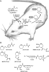
Fig. 1.
Fig. 1.
Cu-free click chemistry in mice. (A) Mice were injected with Ac4ManNAz once daily for 1 wk to allow for metabolic labeling of glycans with SiaNAz. The mice were then injected with a cyclooctyne-FLAG conjugate for in vivo covalent labeling of azido glycans. (B) Panel of FLAG conjugates used in this study.
Fig. 2.
Fig. 2.
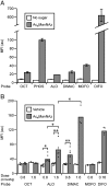
Chemical tagging of azido glycans in vitro and in vivo with cyclooctyne probes. (A) Jurkat cells were incubated with (Gray Bars) or without (White Bars) Ac4ManNAz (25 μM) for 3 d. The cells were then labeled with a FLAG conjugate [250 μM of OCT-FLAG (OCT), PHOS-FLAG (PHOS), ALO-FLAG (ALO), DIMAC-FLAG (DIMAC), MOFO-FLAG (MOFO), or DIFO-FLAG (DIFO)] for 1 h at RT. The cells were stained with FITC-conjugated anti-FLAG antibody (FITC-anti-FLAG) and then analyzed by flow cytometry. Error bars represent the standard deviation of the average of three replicate samples. (B) Mice were injected with either Ac4ManNAz (300 mg/kg, i.p., Gray Bars) or vehicle (70% DMSO, White Bars) once daily for 7 d. On day 8, the mice were injected i.p. with various doses (as indicated) of either OCT-FLAG (OCT), ALO-FLAG (ALO), DIMAC-FLAG (DIMAC), MOFO-FLAG (MOFO), or DIFO-FLAG (DIFO). After 3 h, the mice were euthanized, and splenocytes were isolated, labeled with FITC-anti-FLAG, and analyzed by flow cytometry. Each point represents the average mean fluorescence intensity (MFI) value of three replicate samples from an individual mouse. Each bar represents the average MFI value of splenocytes isolated from separate mice (n = 3–11). MFI is in arbitrary units (au). For all in vivo experiments, ex vivo reactions of isolated splenocytes with FLAG conjugates verified the presence of cell-surface azides for all of these probes (SI Text). ∗ P < 0.02; ∗∗ P < 0.008.
Fig. 3.
Fig. 3.
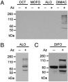
Chemical tagging of serum glycoproteins in vivo using Cu-free click chemistry. Mice were injected with either Ac4ManNAz (300 mg/kg, i.p., +Az) or vehicle (70% DMSO, -Az) once daily for 7 d. On day 8, the mice were injected i.p. with various doses of a cyclooctyne-FLAG probe. Serum was collected and analyzed by Western blot with a horseradish peroxidase-anti-FLAG antibody conjugate (HRP-anti-FLAG). (A) Samples from mice injected with 0.8 mmol/kg OCT-FLAG (OCT), MOFO-FLAG (MOFO), ALO-FLAG (ALO), DIMAC-FLAG (DIMAC); blot was exposed for 3 s. (B) Same blot as (A) exposed for 30 s, showing serum glycoproteins labeled with ALO-FLAG. (C) Samples from mice injected with 0.16 mmol/kg DIFO-FLAG; exposed for 2 s.
Fig. 4.
Fig. 4.
Comparison of Cu-free click chemistry and the Staudinger ligation for labeling splenocyte cell-surface azides in vivo. Mice were injected with Ac4ManNAz (300 mg/kg, i.p., Gray Bars) or vehicle (70% DMSO, White Bars) once daily for 7 d. On day 8, the mice were injected with either (A) DIMAC-FLAG (DIMAC, 0.8 mmol/kg, i.p.), (B) DIFO-FLAG (DIFO, 0.16 mmol/kg, i.p.), or PHOS-FLAG (PHOS) at the same dose. Three h postinjection of the FLAG conjugates, the mice were euthanized, and the splenocytes were harvested, labeled with FITC-anti-FLAG, and analyzed by flow cytometry. Each point represents the average MFI value of three replicate samples from an individual mouse. Each bar represents the average MFI value of splenocytes isolated from separate mice (n = 3–11). ∗ P < 0.02; ∗∗ P < 0.008.
Fig. 5.
Fig. 5.
Cu-free click chemistry and Staudinger ligation products are observed in a variety of tissues in vivo. Mice were injected with Ac4ManNAz (300 mg/kg, i.p., +Az) or vehicle (70% DMSO,-Az) once daily for 7 d. On day 8, the mice were injected with either (A) ALO-FLAG (0.8 mmol/kg, i.p.), blot was exposed overnight; (B) DIMAC-FLAG (0.8 mmol/kg, i.p.), blot was exposed for 5 min; (C) DIFO-FLAG (0.16 mmol/kg, i.p.), blot was exposed for 1 h; or (D) PHOS-FLAG (0.8 mmol/kg, i.p.), blot was exposed for 10 s. Three h postinjection of the FLAG conjugates, the mice were euthanized, and organs (liver, heart, and intestines) were harvested and homogenized. The organ lysates were analyzed by Western blot probing with HRP-anti-FLAG. Each lane represents organ lysate from a single representative mouse.
Fig. 6.
Fig. 6.
DIFO-FLAG binds mouse serum albumin (MSA). Liver lysates from mice injected with Ac4ManNAz (300 mg/kg, i.p., +Az) or vehicle (70% DMSO,-Az) once daily for 7 d, followed by one bolus of DIFO-FLAG (0.16 mmol/kg, i.p.) on day 8, were immunoprecipitated with (AB) an anti-FLAG antibody (FLAG) or an isotype control (iso), or (CD) an anti-MSA antibody (MSA) or an isotype control (iso). The samples were analyzed by Western blot probing for (A) and (C) FLAG or (B) and (D) MSA.

References

    1. Peters LL, et al. The mouse as a model for human biology: A resource guide for complex trait analysis. Nat Rev Genet. 2007;8:58–69. - PubMed
    1. Ohtsubo K, Marth JD. Glycosylation in cellular mechanisms of health and disease. Cell. 2006;126:855–867. - PubMed
    1. Ohtsubo K, et al. Dietary and genetic control of glucose transporter 2 glycosylation promotes insulin secretion in suppressing diabetes. Cell. 2005;123:1307–1321. - PubMed
    1. Hart GW, Housley MP, Slawson C. Cycling of O-linked beta-N-acetylglucosamine on nucleocytoplasmic proteins. Nature. 2007;446:1017–1022. - PubMed
    1. Kayser H, et al. Biosynthesis of a nonphysiological sialic acid in different rat organs, using N-propanoyl-D-hexosamines as precursors. J Biol Chem. 1992;267:16934–16938. - PubMed

Publication types

MeSH terms