. 2010 Jan 26;107(4):1524-8.
doi: 10.1073/pnas.0910341107. Epub 2010 Jan 4.

Dual function of CTLA-4 in regulatory T cells and conventional T cells to prevent multiorgan autoimmunity

Affiliations

Dual function of CTLA-4 in regulatory T cells and conventional T cells to prevent multiorgan autoimmunity

Nitya Jain et al. Proc Natl Acad Sci U S A. .

Abstract

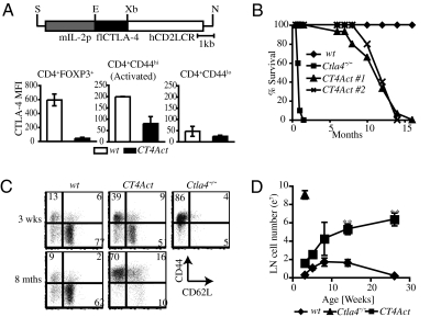
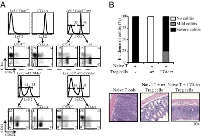
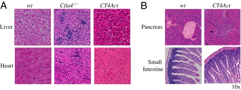
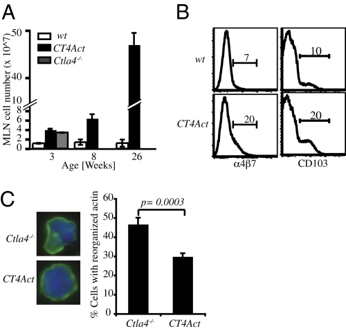
Cytotoxic T lymphocyte antigen-4 (CTLA-4) is an inhibitory receptor on T cells essential for maintaining T cell homeostasis and tolerance to self. Mice lacking CTLA-4 develop an early onset, fatal breakdown in T cell tolerance. Whether this autoimmune disease occurs because of the loss of CTLA-4 function in regulatory T cells, conventional T cells, or both is unclear. We show here that lack of CTLA-4 in regulatory T cells leads to aberrant activation and expansion of conventional T cells. However, CTLA-4 expression in conventional T cells prevents aberrantly activated T cells from infiltrating and fatally damaging nonlymphoid tissues. These results demonstrate that CTLA-4 has a dual function in maintaining T cell tolerance: CTLA-4 in regulatory T cells inhibits inappropriate naïve T cell activation and CTLA-4 in conventional T cells prevents the harmful accumulation of self-reactive pathogenic T cells in vital organs.


Conflict of interest statement

The authors declare no conflict of interest.

Figures

Fig. 1.
Fig. 1.
Massive T cell activation and expansion in mice lacking CTLA-4 expression in Treg cells (A Upper) Restriction enzyme map of Il2pCtla4Tg construct. Full length (fl) CTLA-4 cDNA was cloned 3′ of the mouse Il2 promoter and 5′ of the human CD2 locus-control region (LCR). Restriction endonuclease sites S: Sal1, E: EcoR1, Xb: Xba1, N: Not1. (Lower) Intracellular CTLA-4 expression in T cell subsets from 6- to 12-week-old WT and CT4Act mice was determined by flow cytometry. Data are the average of Mean Fluorescence Intensity (MFI) of CTLA-4 expression determined over at least five independent experiments with two to three mice in each group per experiment. For comparison, background CTLA-4 staining from Ctla4−/− CD4+T cells had MFI of 30. (B) WT (n = 10, ◆), Ctla4−/− (n = 10, ■ ) and two founder lines of CT4Act (n = 15, ▲, X) mice were observed for signs of disease and mortality over the indicated months. Results are represented as percent survival, which was calculated as 100× (number of surviving mice/total number of mice) at each time point. (C) Representative flow cytometric dot plot showing expression of activation markers CD44 and CD62L on CD4+FOXP3 T cells in peripheral LNs of WT and CT4Act mice at 3 weeks and 8 months of age and Ctla4−/− mice at 3 weeks of age. Data are representative of at least three independent experiments with two to three mice per group. (D) Total cellularity of inguinal, axillary and brachial LNs in WT (◆), Ctla4−/− (▲), and CT4Act (■) mice at different ages. Data are a mean of at least four mice per group at each time point. (** P ≤ 0.001)
Fig. 2.
Fig. 2.
Ctla4-deficient FOXP3+Treg cells are functionally impaired in vivo (A) BMCs were generated in Rag1−/− mice by injecting T cell-depleted BM cells from either Ly5.1+ Ctla4−/− or CT4Act mice (Upper Left, negative controls), or coinjecting a mix of BM cells from WT and Ly5.1+ Ctla4−/− mice (Upper Right), Ly5.1 WT and CT4Act mice (Lower Left), and Ly5.1 Ctla4−/− and CT4Act mice (Lower Right). Six to eight weeks after reconstitution, before some BMCs became fatally sick, mice were euthanized and LN cells were stained with Ly5 congenic marker to determine contributions of each group to the peripheral lymphocyte compartment. Ly5.2+ and Ly5.2 CD4+ T cells were further analyzed for the expression of activation markers CD44 and CD62L. Data are representative of two independent experiments with three mice in each group. (B) CD4+CD25+ Treg cells from WT, and CT4Act mice were transferred with WT CD4+CD25neg T cells into Rag1−/− mice to test their ability to prevent colitis. (Upper) Colitis was scored as described in ref. . Data are representative of two independent experiments with four to five mice in each group. (Lower) Representative H&E stained sections of colons of Rag1−/− mice after transfer of T cells. Mice that received naïve WT T cells (Left) developed severe colitis, which was prevented by cotransfer of WT Treg cells (Middle). Mice constituted with naïve WT T cells and CT4Act Treg cells (Right) developed colitis, displaying substantial inflammatory infiltrates and significant epithelial hyperplasia with loss of goblet cells. However, the severity of colitis was milder as compared to mice receiving only naive WT T cells. (Original magnification, ×10.)
Fig. 3.
Fig. 3.
CTLA-4 expression in activated T cells restricts tissue infiltration and pathology. Lymphocyte infiltration in WT (7 months old), Ctla4−/− (3-4 weeks old) and CT4Act (7 months old) mice was determined by histology. (A) Representative H&E stained sections of liver and heart showed extensive lymphocyte infiltration only in sick Ctla4−/− mice. (B) Pancreatic β-islets and small intestines were infiltrated with lymphocytes in 8-month-old CT4Act mice. At least two sections/mouse and multiple fields were analyzed. (Original magnification, ×10.)
Fig. 4.
Fig. 4.
Altered homing capabilities of CTLA-4 expressing activated T cells in CT4Act mice. (A) Total cellularity of MLNs at different ages in WT, Ctla4−/− and CT4Act mice. Data are a mean of at least four mice per group at each time point. (B) Expression of homing markers α4β7 and CD103 on CD4+CD44hi activated T cells from MLN of 2- to 3-month-old WT and CT4Act mice as determined by flow cytometry. Data are representative of three independent experiments with three to four mice in each group. (C Left) Representative F-actin localizations in CD4+ T cells from MLNs of Ctla4−/− and CT4Act mice analyzed by fluorescence microscopy. Green: F-actin staining using phalloidin-Alexa 488; Blue: nuclear staining using DAPI. (Right) Frequency of CD4+T cells with polarized F-actin. Data are generated from three independent experiments with a total of at least 400 cells analyzed for each genotype.

Similar articles

Cited by

References

    1. Waterhouse P, et al. Lymphoproliferative disorders with early lethality in mice deficient in CTLA-4. Science. 1995;270:985–988. - PubMed
    1. Tivol EA, et al. Loss of CTLA-4 leads to massive lymphoproliferation and fatal multiorgan tissue destruction, revealing a critical negative regulatory role of CTLA-4. Immunity. 1995;3:541–547. - PubMed
    1. Chambers CA, Sullivan TJ, Allison JP. Lymphoproliferation in CTLA-4-deficient mice is mediated by costimulation-dependent activation of CD4+ T cells. Immunity. 1997;7:885–895. - PubMed
    1. Perkins D, et al. Regulation of CTLA-4 expression during T cell activation. J Immunol. 1996;156:4154–4159. - PubMed
    1. Takahashi T, et al. Immunologic self-tolerance maintained by CD25(+)CD4(+) regulatory T cells constitutively expressing cytotoxic T lymphocyte-associated antigen 4. J Exp Med. 2000;192:303–310. - PMC - PubMed

Publication types