. 2010 Jan 26;107(4):1321-6.
doi: 10.1073/pnas.0907304107. Epub 2010 Jan 11.

Detecting and understanding combinatorial mutation patterns responsible for HIV drug resistance

Affiliations

Detecting and understanding combinatorial mutation patterns responsible for HIV drug resistance

Jing Zhang et al. Proc Natl Acad Sci U S A. .

Abstract

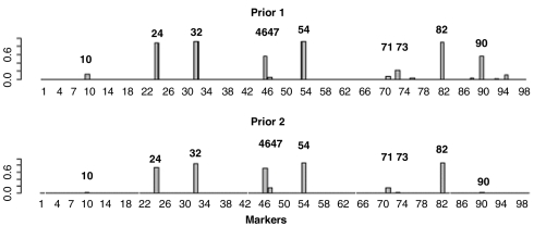
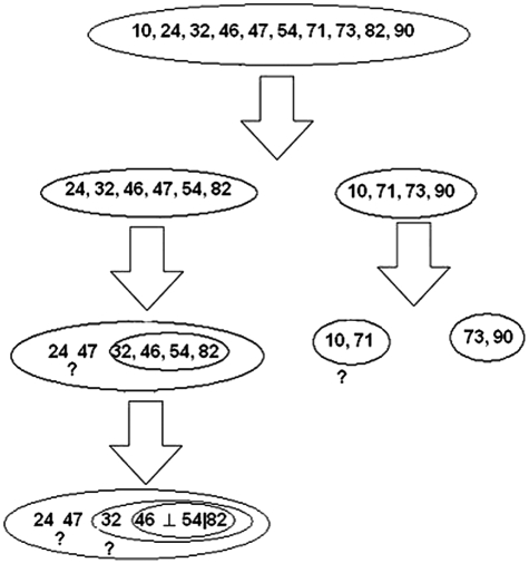
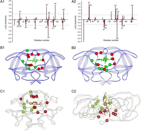
We propose a systematic approach for a better understanding of how HIV viruses employ various combinations of mutations to resist drug treatments, which is critical to developing new drugs and optimizing the use of existing drugs. By probabilistically modeling mutations in the HIV-1 protease or reverse transcriptase (RT) isolated from drug-treated patients, we present a statistical procedure that first detects mutation combinations associated with drug resistance and then infers detailed interaction structures of these mutations. The molecular basis of our statistical predictions is further studied by using molecular dynamics simulations and free energy calculations. We have demonstrated the usefulness of this systematic procedure on three HIV drugs, (Indinavir, Zidovudine, and Nevirapine), discovered unique interaction features between viral mutations induced by these drugs, and revealed the structural basis of such interactions.


Conflict of interest statement

The authors declare no conflict of interest.

Figures

Fig. 1.
Fig. 1.
The posterior probabilities for each mutation to be associated interactively with indinavir treatment. The Upper shows the posterior probabilities using prior one, which assumes that it is equally likely (1/3) for a mutation to be unassociated, individually associated, and interactively associated with the drug treatment. The Lower shows the posterior probabilities using a more stringent prior (prior two) assuming that only two makers are expected to be associated with the drug, either individually or interactively.
Fig. 2.
Fig. 2.
Detection of a detailed mutation interaction structure for resisting indinavir. Positions 46 and 54 are conditionally independent given position 82, denoted as 42⊥54|82. The ? indicates where we are not able to confidently infer the dependence structure (SI Text).
Fig. 3.
Fig. 3.
Energetic and structural insight of the resistance mechanism. A1: The difference between each residue’s contribution to the interaction with indinavir in (A1) the M46I/I54V and the M46I; (A2) the I54V/V82A and the V82A. ΔΔG was calculated by subtracting each residue’s interaction energy in the single mutant (e.g., M46I) from the double mutant (M46I/I54V). Residues with absolute value greater than 0.75 kcal/mol are labeled. Structural distributions of important residues in Fig 3 A1 and A2 are shown in B1 and B2, resp. The protease is shown in Blue Strand and indinavir in Green Stick. Residues with negative and positive ΔΔG’s, which represent residues contributing more and less favorably to binding with indinavir in the double mutant (e.g., M46I/I54V) than in the single mutant (e.g., M46I) resp., are shown in Red and Green CPK models, resp. The favorable residues to the binding of indinavir to the M46I/54V mutant are shown as the Red CPK model and those of the unfavorable residues as the Green CPK model. Alignment of the average structure of the double and single mutant complexes (C1) between M46I/54V and M46I mutated; (C2) between I54V/V82A and V82A. The average structure was obtained by averaging the 125 snapshots taken from 0.5–3.0 ns MD simulations. The double (e.g., M46I/I54V) and single (e.g., M46I) protease mutants are shown in Blue and Green strands, resp. Indinavirs bound to the double (e.g., M46I/I54V) and single (e.g., M46I) are shown in Red and Green Sticks, resp. The Pink Arrow shows the configurational change of indinavir in the two complexes. The cooperation between, for example, V54 and A82 significantly changes the active site’s conformation that further enhances resistance caused by the mutation at position 82 alone. The conformational change is manifested in the alignment of the average structures of the double (e.g., I54V/V82A) and single (e.g., V82A) mutant complexes.

References

    1. Lengauer T, Sander O, Sierra S, Thielen A, Kaiser R. Bioinformatics prediction of HIV coreceptor usage. Nat Biotechnol. 2007;25:1407–1410. - PubMed
    1. Lengauer T, Sing L. Bioinformatics-assisted anti-HIV therapy. Nat Rev Microbiol. 2006;4:790–797. - PubMed
    1. Shafer RW. Genotypic testing for Human Immunodeficiency Virus type 1 drug resistance. Clin Microbiol Rev. 2002;15:247–277. - PMC - PubMed
    1. Liu TF, Shafer RW. Web resources for HIV type 1 genotypic-resistance test interpretation. Clin Infect Diseases. 2006;42:1608–1618. - PMC - PubMed
    1. Johnson VA, et al. Update of the drug resistance mutations in HIV-1: Spring 2008. Top HIV Med. 2008;16:62–68. - PubMed

Publication types

MeSH terms