. 2010 Feb 2;107(5):1906-11.
doi: 10.1073/pnas.0914052107. Epub 2010 Jan 11.

Direct contacts between extracellular membrane-proximal domains are required for VEGF receptor activation and cell signaling

Affiliations

Direct contacts between extracellular membrane-proximal domains are required for VEGF receptor activation and cell signaling

Yan Yang et al. Proc Natl Acad Sci U S A. .

Abstract

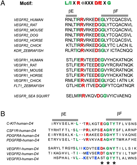
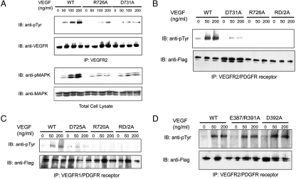
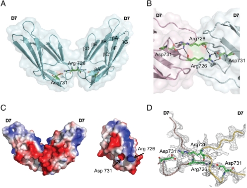
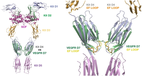
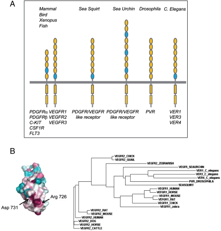
Structural analyses of the extracellular region of stem cell factor (SCF) receptor (also designated KIT) in complex with SCF revealed a sequence motif in a loop in the fourth Ig-like domain (D4) that is responsible for forming homotypic receptor contacts and for ligand-induced KIT activation and cell signaling. An identical motif was identified in the most membrane-proximal seventh Ig-like domain (D7) of vascular endothelial growth factor receptor 1 (VEGFR1), VEGFR2, and VEGFR3. In this report we demonstrate that ligand-induced tyrosine autophosphorylation and cell signaling via VEGFR1 or VEGFR2 harboring mutations in critical residues (Arg726 or Asp731) in D7 are strongly impaired. We also describe the crystal structure of D7 of VEGFR2 to a resolution of 2.7 A. The structure shows that homotypic D7 contacts are mediated by salt bridges and van der Waals contacts formed between Arg726 of one protomer and Asp731 of the other protomer. The structure of D7 dimer is very similar to the structure of D4 dimers seen in the crystal structure of KIT extracellular region in complex with SCF. The high similarity between VEGFR D7 and KIT D4 in both structure and function provides further evidence for common ancestral origins of type III and type V RTKs. It also reveals a conserved mechanism for RTK activation and a novel target for pharmacological intervention of pathologically activated RTKs.


Conflict of interest statement

The authors declare no conflict of interest.

Figures

Fig. 1.
Fig. 1.
Amino Acid sequence analysis of D7 and D4 of VEGFR2 extracellular region. (A) Structure-based multiple sequence alignment of predicted EF-loop region of D7 of VEGFR1 and VEGFR2 from different species. Key amino acids in the I-set Ig frame are highlighted in green, and the conserved Arg/Asp pair in the EF loop is highlighted in red. (B) Comparison of predicted EF-loop region of D4 from VEGFR and D4 of KIT, CSF1R, and PDGFRs (type-III RTK). Key amino acids in the I-set Ig frame are highlighted in green, and the conserved Arg/Asp or Glu pair in the EF loop is highlighted in red. Nonconserved amino acids with opposite charge in the EF loop are highlighted in blue. The conserved Y-corner motif is marked with an asterisk (*).
Fig. 2.
Fig. 2.
Ligand-induced activation of VEGFR2 is compromised by mutations in EF-loop region of D7 but not affected by mutation in EF-loop region of D4. (A) HEK293 cells transiently expressing WT VEGFR2, the R726A, or the D731A VEGFR2 mutants were stimulated with indicated amount of VEGF-A for 5 min at 37 °C. Lysates from unstimulated or VEGF-A–stimulated cells were subjected to immunoprecipitation with anti-VEGFR2 antibodies followed by immunoblotting (IB) with anti-pTyr, or with anti-VEGFR2 antibodies. Total cell lysate from the same experiment was analyzed by SDS-PAGE followed by immunoblotting with anti-phosphoMAPK (pMAPK) or anti-MAPK antibodies. (B) Serum-starved 3T3 cells stably expressing a VEGFR2-PDGFRβ chimeric receptor or chimeric receptors harboring mutations in D7 region (R726A, D731A, or an R726/D731 double mutation designated RD/2A) were stimulated with VEGF-A for 5 min at 37 °C. Lysates from unstimulated or VEGF-A–stimulated cells were subjected to immunoprecipitation with antibodies against the cytoplasmic region of the chimeric receptor followed by immunoblotting with either anti-pTyr or anti-tag (FLAG) antibodies, respectively. (C) Serum-starved 3T3 cells stably expressing a VEGFR1-PDGFR chimeric receptor or chimeric receptors harboring mutation in the D7 region (R720A, D725A, or R720D725/2A double mutations) were analyzed as described in (B). (D) 3T3 cells expressing a VEGFR2-PDGFR chimeric receptor or chimeric receptors harboring mutations in D4 region (D392A or E387/R391A double mutations) were analyzed as described in (B).
Fig. 3.
Fig. 3.
Structure of VEGFR2 D7 homodimer. (A) A ribbon diagram and a transparent molecular surface of D7 homodimer structure (side view). Asp731 and Arg726 are shown as a stick model. (B) A close view of the homotypic D7 interface of the two neighboring molecules (pink and green). Salt bridges formed between Asp731 and Arg726 are shown as dashed lines. (C) Charge distribution of D7 homodimer (side view) is shown as a surface potential model (Left). View of D7 surface that mediates homotypic contacts (Right). (D) 2Fo-Fc electron density map contoured at 1.1σ level showing a view of the D7–D7 interface. The backbones of VEGFR D7 protomers are represented as pink and yellow tubes, respectively.
Fig. 4.
Fig. 4.
Superposition of the structure of D7 of VEGFR2 with the structure of D4 of dimeric KIT-SCF complex. Overlay of VEGFR D7 structure (PDB ID code: 3KVQ) and the structure of KIT dimer in complex with SCF (PDB ID code: 2E9W) (Left). A closer view of superimposed D7 and D4 regions reveals high similarity in domain arrangement and homotypic contacts (Right). VEGFR2 D7 is illustrated in green and the EF loop is in yellow. D4 of KIT is illustrated in gray and its EF loop is in orange.
Fig. 5.
Fig. 5.
Phylogenetic analysis of VEGFR1 and VEGFR2. (A) Location of the conserved EF loop in type-III and type-V RTKs from various species. Ig-like domains containing a conserved EF-loop motif are marked in blue. (B) Color-coded conservation pattern of VEGFR2 D7 region. Amino acid sequences of human VEGFR2 were used as query to search nonredundant database for homologous sequences, using PSI-BLAST. Sequence alignment of D7 was performed using ClustalW, manually adjusted based on the IgSF fold restrains for 20 key residues. The alignment of amino acid sequences was submitted to the Consurf 3.0 server to generate maximum-likelihood normalized evolutionary rates for each position. Cyan through maroon is used for labeling from variable to conserved amino acids. Phylogenetic tree of VEGFR1 and VEGFR2 are generated by the neighboring-joining method based using Clustal W2.

References

    1. Olsson A-K, Dimberg A, Kreuger J, Claesson-Welsh L. VEGF receptor signalling in control of vascular function. Nat Rev Mol Cell Biol. 2006;7(5):359–371. - PubMed
    1. Barleon B, et al. Mapping of the sites for ligand binding and receptor dimerization at the extracellular domain of the vascular endothelial growth factor receptor FLT-1. J Biol Chem. 1997;272(16):10382–10388. - PubMed
    1. Shinkai A, et al. Mapping of the sites involved in ligand association and dissociation at the extracellular domain of the kinase insert domain-containing receptor for vascular endothelial growth factor. J Biol Chem. 1998;273(47):31283–31288. - PubMed
    1. Wiesmann C, et al. Crystal structure at 1.7 A resolution of VEGF in complex with domain 2 of the Flt-1 receptor. Cell. 1997;91(5):695–704. - PubMed
    1. Shibuya M, Claesson-Welsh L. Signal transduction by VEGF receptors in regulation of angiogenesis and lymphangiogenesis. Exp Cell Res. 2006;312(5):549–560. (in eng) - PubMed

Publication types

MeSH terms

Substances