. 2010 Jan 26;107(4):1437-42.
doi: 10.1073/pnas.0911427107. Epub 2010 Jan 4.

Hippo signaling is a potent in vivo growth and tumor suppressor pathway in the mammalian liver

Affiliations

Hippo signaling is a potent in vivo growth and tumor suppressor pathway in the mammalian liver

Li Lu et al. Proc Natl Acad Sci U S A. .

Abstract

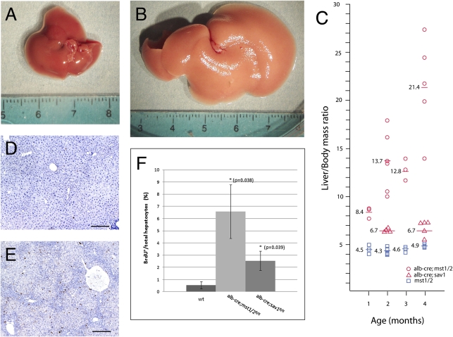
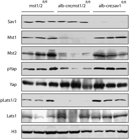
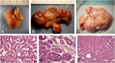
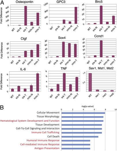
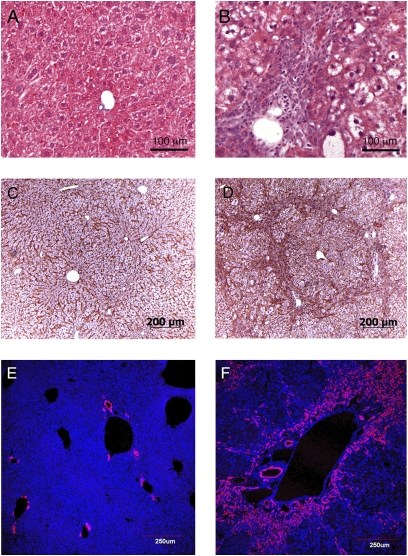
How organ size is controlled in mammals is not currently understood. In Drosophila the Hippo signaling pathway functions to suppress growth in imaginal discs and has been suggested to control organ size. To investigate the role of hippo signaling in regulation of mammalian organ size we have generated conditional alleles of Sav1, mst1, and mst2, orthologs of Drosophila Salvador and hippo, respectively. Specific deletion of both mst1 and mst2 in hepatocytes results in significantly enlarged livers due to excessive proliferation. By the age of 5-6 months, mst1/2 conditional mutant livers have multiple foci of liver tumors, indicating that the combined activities of mst1 and mst2 act as redundant tumor suppressors in hepatocytes. Similar findings were obtained with liver-specific deletion of Sav1, a second core Hippo signaling component that facilitates activation of mst1 and mst2. Tumors from sav1 mutants exhibited varied morphology, suggesting a mixed-lineage origin of tumor-initiating cells. Transcriptional profiling of liver tissues from both mst1/2 and sav1 conditional mutants revealed a network of Hippo signaling regulated genes with specific enrichment for genes involved in immune and inflammatory responses. Histological and immunological characterization of mst1/2 double mutant liver tissues revealed abundant accumulation of adult facultative stem cells termed oval cells in periductal regions. Because oval cells induction is commonly associated with liver injury and tumor formation, it is likely that these cells contribute to the enlarged livers and hepatomas that we observe in sav1 and mst1/2 mutants. Taken together, our results demonstrate that the Hippo signaling pathway is a critical regulator of mammalian liver growth and a potent suppressor of liver tumor formation.


Conflict of interest statement

The authors declare no conflict of interest.

Figures

Fig. 1.
Fig. 1.
Mst1 and mst2 are required to restrict liver growth and to maintain hepatocyte quiescence. In contrast to wild type (A), albumin-cre;mst1/2 double mutants (B) have significantly enlarged livers at 2 months of age. (C) A plot of liver/body as a function of age shows increased liver size of mst1/2 mutants at 1 month. Continued growth is evident at 2 and 3 months of age. Modest increases in albumin-cre;sav1 mutant liver sizes are seen at 2 and 4 months of age. Few wild-type hepatocytes incorporate BrdU at 2 months of age (D), although albumin-cre;mst1/2 mutants show abundant BrdU incorporation (E). (F) Quantification of BrdU labeling. Albumin -cre;mst1/2 double mutants and albumin-cre;sav1 mutants exhibit 12-fold and 5-fold increases in BrdU incorporation respectively.
Fig. 2.
Fig. 2.
Western analysis of albumin-cre;sav1 and albumin-cre;mst1/2 double mutant tissues. Cells enriched for hepatocytes show loss of sav1 protein in albumin-cre;sav1 mutants and reduced amounts of mst1 and mst2 proteins in albumin-cre;mst1/2 mutants. Each lane represents proteins extracted from independent mutant livers. Phosphorylation of Yap and Lats is reduced in albumin-cre;mst1/2 double mutant hepatocytes but not in albumin-cre;sav1 mutant cells. Histone H3 (H3) was used as a loading control.
Fig. 3.
Fig. 3.
Hepatoma formation in Hippo pathway mutants. Wild-type livers display a normal appearance, devoid of tumor foci (A). In contrast, conditional deletion of sav1 in hepatocytes results in large, multifocal tumors (B). Likewise, albumin-cre;mst1/2 mutant livers are significantly enlarged relative to wild type and display multiple focal tumor nodules (C). Histological examination (D and E) reveals both well and poorly differentiated hepatocellular carcinoma (D and F) and cholangiocarcinoma (E).
Fig. 4.
Fig. 4.
Transcriptional analysis of sav1 and mst1/2 mutant liver tissue. (A) Quantitative RT-PCR analysis of gpc3, osteopontin, ctgf, sox4, survivin, cyclin D1, IL-6, and TNF-α validates and extends the microarray data indicating differential expression of these transcripts. Reduction of sav1, mst1, and mst2 transcript levels is readily apparent in hepatocyte-enriched cell fractions from albumin-cre;sav1 and albumin-cre;mst1/2 mutants. (B) Gene set enrichment analysis reveals overrepresentation of transcripts involved in immune response in Hippo pathway mutant tissues.
Fig. 5.
Fig. 5.
Histology and oval cell response in albumin-cre;mst1/2 double mutants are consistent with chronic liver injury. The normal liver (A) is composed of regularly spaced plates of hepatocytes surrounding the portal vein. In contrast, albuin-cre;mst1/2 mutants display a disorganized arrangements of hepatocytes, many of which have an abnormal clear cytoplasm with H&E stain. Many small periductal cells are visible that are not present in wild-type liver sections. Overall hepatocyte disorganization and infiltrating periductal cells are prominently visualized with cell membrane β-catenin staining of wild type (C) versus mutant (D). The A6 antibody, a marker for oval and ductal cells, stains only ductal cells in wild-type (E) tissues, but stains both ductal cells and periductal cells in albumin-cre;mst1/2 double mutants (F).

Similar articles

Cited by

References

    1. Edgar BA. From cell structure to transcription: Hippo forges a new path. Cell. 2006;124:267–273. - PubMed
    1. Harvey K, Tapon N. The Salvador-Warts-Hippo pathway - an emerging tumour-suppressor network. Nat Rev Cancer. 2007;7:182–191. - PubMed
    1. Zeng Q, Hong W. The emerging role of the hippo pathway in cell contact inhibition, organ size control, and cancer development in mammals. Cancer Cell. 2008;13:188–192. - PubMed
    1. Zhao B, Lei QY, Guan KL. The Hippo-YAP pathway: New connections between regulation of organ size and cancer. Curr Opin Cell Biol. 2008;20:638–646. - PMC - PubMed
    1. St John MA, et al. Mice deficient of Lats1 develop soft-tissue sarcomas, ovarian tumours and pituitary dysfunction. Nat Genet. 1999;21:182–186. - PubMed

Publication types

MeSH terms