

Computational and single-molecule force studies of a macro domain protein reveal a key molecular determinant for mechanical stability
- PMID: 20080695
- PMCID: PMC2836608
- DOI: 10.1073/pnas.0905796107
Computational and single-molecule force studies of a macro domain protein reveal a key molecular determinant for mechanical stability
Abstract
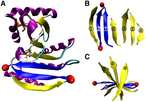
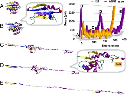
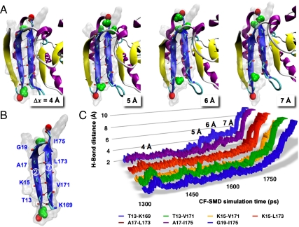
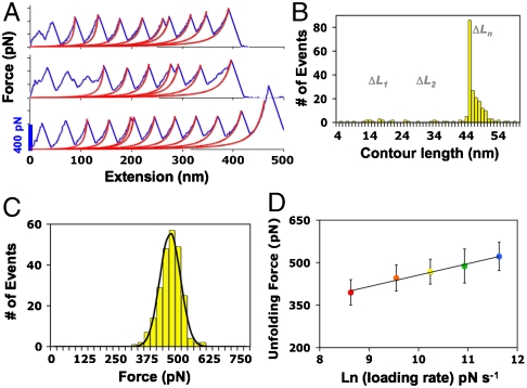
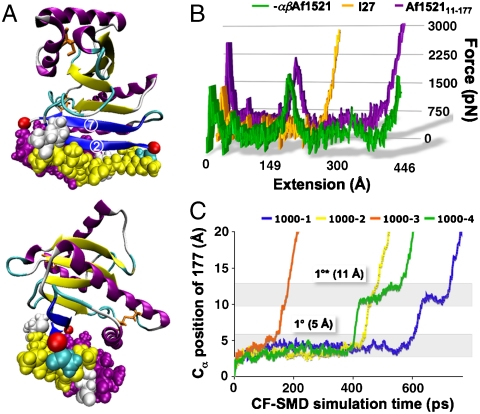
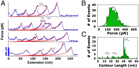
Resolving molecular determinants of mechanical stability of proteins is crucial in the rational design of advanced biomaterials for use in biomedical and nanotechnological applications. Here we present an interdisciplinary study combining bioinformatics screening, steered molecular dynamics simulations, protein engineering, and single-molecule force spectroscopy that explores the mechanical properties of a macro domain protein with mixed alpha + beta topology. The unique architecture is defined by a single seven-stranded beta-sheet in the core of the protein flanked by five alpha-helices. Unlike mechanically stable proteins studied thus far, the macro domain provides the distinct advantage of having the key load-bearing hydrogen bonds (H bonds) buried in the hydrophobic core protected from water attacks. This feature allows direct measurement of the force required to break apart the load-bearing H bonds under locally hydrophobic conditions. Steered molecular dynamics simulations predicted extremely high mechanical stability of the macro domain by using constant velocity and constant force methods. Single-molecule force spectroscopy experiments confirm the exceptional mechanical strength of the macro domain, measuring a rupture force as high as 570 pN. Furthermore, through selective deletion of shielding peptide segments, we examined the same key H bonds under hydrophilic environments in which the beta-strands are exposed to solvent and verify that the high mechanical stability of the macro domain results from excellent shielding of the load-bearing H bonds from competing water. Our study reveals that shielding water accessibility to the load-bearing strands is a critical molecular determinant for enhancing the mechanical stability of proteins.
Conflict of interest statement
The authors declare no conflict of interest.
Figures
Similar articles
-
Single-molecule force spectroscopy reveals a mechanically stable protein fold and the rational tuning of its mechanical stability.Proc Natl Acad Sci U S A. 2007 May 29;104(22):9278-83. doi: 10.1073/pnas.0700351104. Epub 2007 May 21. Proc Natl Acad Sci U S A. 2007. PMID: 17517616 Free PMC article.
-
Stabilization provided by neighboring strands is critical for the mechanical stability of proteins.Biophys J. 2008 Oct;95(8):3935-42. doi: 10.1529/biophysj.108.134072. Epub 2008 Jul 3. Biophys J. 2008. PMID: 18599623 Free PMC article.
-
Single molecule force spectroscopy reveals critical roles of hydrophobic core packing in determining the mechanical stability of protein GB1.Langmuir. 2012 Aug 21;28(33):12319-25. doi: 10.1021/la301940g. Epub 2012 Aug 6. Langmuir. 2012. PMID: 22823458
-
Mechanical design of proteins studied by single-molecule force spectroscopy and protein engineering.Prog Biophys Mol Biol. 2000;74(1-2):63-91. doi: 10.1016/s0079-6107(00)00017-1. Prog Biophys Mol Biol. 2000. PMID: 11106807 Review.
-
Probing the mechanical stability of proteins using the atomic force microscope.Biochem Soc Trans. 2007 Dec;35(Pt 6):1564-8. doi: 10.1042/BST0351564. Biochem Soc Trans. 2007. PMID: 18031267 Review.
Cited by
-
Protein folding at single-molecule resolution.Biochim Biophys Acta. 2011 Aug;1814(8):1021-9. doi: 10.1016/j.bbapap.2011.01.011. Epub 2011 Feb 17. Biochim Biophys Acta. 2011. PMID: 21303706 Free PMC article. Review.
-
Engineering proteins with enhanced mechanical stability by force-specific sequence motifs.Proteins. 2012 May;80(5):1308-15. doi: 10.1002/prot.24027. Epub 2012 Feb 10. Proteins. 2012. PMID: 22274941 Free PMC article.
-
The structure of misfolded amyloidogenic dimers: computational analysis of force spectroscopy data.Biophys J. 2014 Dec 16;107(12):2903-2910. doi: 10.1016/j.bpj.2014.10.053. Biophys J. 2014. PMID: 25517155 Free PMC article.
-
The Gearbox of the Bacterial Flagellar Motor Switch.Structure. 2016 Jul 6;24(7):1209-20. doi: 10.1016/j.str.2016.05.012. Epub 2016 Jun 23. Structure. 2016. PMID: 27345932 Free PMC article.
-
Contribution of hydrophobic interactions to protein mechanical stability.Comput Struct Biotechnol J. 2022 Apr 21;20:1946-1956. doi: 10.1016/j.csbj.2022.04.025. eCollection 2022. Comput Struct Biotechnol J. 2022. PMID: 35521554 Free PMC article.
References
-
- Buehler MJ, Keten S, Ackbarow T. Theoretical and computational hierarchical nanomechanics of protein materials: Deformation and fracture. Prog Mater Sci. 2008;53:1101–1241.
-
- Li H. Mechanical engineering of elastomeric proteins: Toward designing new protein building blocks for biomaterials. Adv Funct Mater. 2008;18:2643–2657.
-
- Goodsell DS. Bionanotechnology: Lessons from Nature. Hoboken, NJ: Wiley–Liss; 2004.
-
- Bao G, Suresh S. Cell and molecular mechanics of biological materials. Nat Mater. 2003;2:715–725. - PubMed
-
- Sotomayor M, Schulten K. Single-molecule experiments in vitro and in silico. Science. 2007;316:1144–1148. - PubMed
Publication types
MeSH terms
Substances
Grants and funding
LinkOut - more resources
Full Text Sources
Other Literature Sources
