. 2010 Feb 2;107(5):2048-53.
doi: 10.1073/pnas.0912704107. Epub 2010 Jan 13.

Progression from a stem cell-like state to early differentiation in the C. elegans germ line

Affiliations

Progression from a stem cell-like state to early differentiation in the C. elegans germ line

Olivier Cinquin et al. Proc Natl Acad Sci U S A. .

Abstract

Controls of stem cell maintenance and early differentiation are known in several systems. However, the progression from stem cell self-renewal to overt signs of early differentiation is a poorly understood but important problem in stem cell biology. The Caenorhabditis elegans germ line provides a genetically defined model for studying that progression. In this system, a single-celled mesenchymal niche, the distal tip cell (DTC), employs GLP-1/Notch signaling and an RNA regulatory network to balance self-renewal and early differentiation within the "mitotic region," which continuously self-renews while generating new gametes. Here, we investigate germ cells in the mitotic region for their capacity to differentiate and their state of maturation. Two distinct pools emerge. The "distal pool" is maintained by the DTC in an essentially uniform and immature or "stem cell-like" state; the "proximal pool," by contrast, contains cells that are maturing toward early differentiation and are likely transit-amplifying cells. A rough estimate of pool sizes is 30-70 germ cells in the distal immature pool and approximately 150 in the proximal transit-amplifying pool. We present a simple model for how the network underlying the switch between self-renewal and early differentiation may be acting in these two pools. According to our model, the self-renewal mode of the network maintains the distal pool in an immature state, whereas the transition between self-renewal and early differentiation modes of the network underlies the graded maturation of germ cells in the proximal pool. We discuss implications of this model for controls of stem cells more broadly.


Conflict of interest statement

The authors declare no conflict of interest.

Figures

Fig. 1.
Fig. 1.
Two pools with distinct differentiation capacities in the mitotic region. (A) Schematic of adult hermaphrodite distal gonad. Red, DTC; yellow circles, germ cells in mitotic cell cycle; *, inferred actual stem cells; green circles, germ cells in meiotic S-phase; green crescents, germ cells in early meiotic prophase. Movement through the mitotic region is inferred by the continuous movement of germ cells from the mitotic region to more proximal regions. Neighboring germ cells are often in distinct phases of the cell cycle. (B, D, and E) Confocal images of adult distal gonads. Dashed line, boundary between mitotic region and early meiotic prophase (the boundary is not always straight; it is drawn as a straight line for simplicity); dotted line, likely position of meiotic S phase; white arrowhead, distal end; white arrows, crescent-shaped DNA typical of meiotic prophase. (B) Wild-type distal gonad, single confocal section. DTC is stained with antibodies to GFP (green), germ-line membranes with antibodies to the GLP-1/Notch receptor (red), and DNA with DAPI (blue). (C) Simplified regulatory network for control of decision between self-renewal or early differentiation (meiotic entry). (D and E) Confocal projections of distal emb-30 gonads at permissive temperature (D) [note that the most proximal PH3 in this germ line is not at the average position (see Fig. 1F)] and 15 h after a shift to restrictive temperature (E). Blue, DAPI-stained DNA; red, GLD-1; green, anti-PH3; small red carat, initial GLD-1 step; large red carat, second GLD-1 step; green arrow, most proximal mitotic division (PH3). D and E each show the same germ line with different fluorophores in Left, Center, and Right. (F) Position of key features in mitotic region with time after emb-30 shift to restrictive temperature. Error bars represent 95% confidence intervals. Black dotted lines mark the rough boundary (6-8 gcd) between the distal and proximal pools. (G) Summary diagrams.
Fig. 2.
Fig. 2.
The DTC maintains the emb-30 distal pool in an immature state. (A) Experimental design. (B-D) Projections of confocal sections. White arrowhead marks distal end of gonad; white arrows mark meiotic prophase nuclei. (B) Unablated emb-30 gonad incubated at 25 °C for 21 h. Nuclei are degenerating. GLD-1 (red); DAPI (cyan). Red carat marks GLD-1 border. (C–E) Ablated emb-30 gonads. (C) GLD-1 (red); DAPI (cyan). (D) Chromosomal HIM-3 (pink); DAPI (blue). (E) EdU (pink, labeled for 12 h at 25 °C after ablation); DAPI (blue). Not all meiotic prophase nuclei contain EdU. Arrowheads: 1, no labeling with EdU; 2, weakly labeled with EdU; 3, well labeled with EdU.
Fig. 3.
Fig. 3.
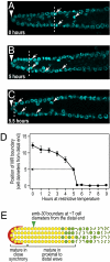
Removal of Notch signaling reveals two pools within the mitotic region. (A-C) Distal gonads from glp-1(ts) adults grown at permissive temperature (A) or at restrictive temperature for the indicated times (B and C). Conventions as in Fig. 1. (D) Graph showing the position of meiotic entry as a function of time after glp-1(ts) adults were shifted to restrictive temperature. Error bars represent 95% median confidence intervals. (E) Summary diagram. The boundary between distal and proximal pools is similar although not identical in glp-1 (brackets) and emb-30 (green arrowhead) experiments (Fig. 1).
Fig. 4.
Fig. 4.
Model for network control of the progression from stem cells through transit-amplifying cells and into early differentiation. (A) Two pools with distinct properties in the mitotic region. Germ cells in the distal pool are maintained in a stem cell–like state; germ cells in the proximal pool are maturing from the stem cell–like state to the early differentiated state. (B–D) Models for network control of the progression from stem cell to early differentiation in three different systems. The network transition between states is drawn as a line for simplicity. SC, stem cell state; Diff, early differentiated state; arrowhead, trigger to leave stem cell state and start network maturation toward the early differentiated state. (B) Model for network control of the C. elegans distal germ line. (C) Model for network control of a stem cell asymmetric division. (D) Model for network control of a vertebrate stem cell system that makes a large number of transit-amplifying cells.

Similar articles

Cited by

References

    1. Li L, Xie T. Stem cell niche: Structure and function. Annu Rev Cell Dev Biol. 2005;21:605–631. - PubMed
    1. Morrison SJ, Spradling AC. Stem cells and niches: Mechanisms that promote stem cell maintenance throughout life. Cell. 2008;132:598–611. - PMC - PubMed
    1. Knoblich JA. Mechanisms of asymmetric stem cell division. Cell. 2008;132:583–597. - PubMed
    1. Morrison SJ, Kimble J. Asymmetric and symmetric stem-cell divisions in development and cancer. Nature. 2006;441:1068–1074. - PubMed
    1. Kimble J, Crittenden SL. Controls of germline stem cells, entry into meiosis, and the sperm/oocyte decision in Caenorhabditis elegans. Annu Rev Cell Dev Biol. 2007;23:405–433. - PubMed

Publication types

MeSH terms

Substances

LinkOut - more resources