. 2010 Jan 19;107(3):1201-6.
doi: 10.1073/pnas.0909015107. Epub 2009 Dec 28.

NeuroD1 induces terminal neuronal differentiation in olfactory neurogenesis

Affiliations

NeuroD1 induces terminal neuronal differentiation in olfactory neurogenesis

Camille Boutin et al. Proc Natl Acad Sci U S A. .

Abstract

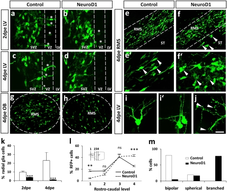
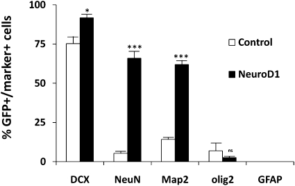
After their generation and specification in periventricular regions, neuronal precursors maintain an immature and migratory state until their arrival in the respective target structures. Only here are terminal differentiation and synaptic integration induced. Although the molecular control of neuronal specification has started to be elucidated, little is known about the factors that control the latest maturation steps. We aimed at identifying factors that induce terminal differentiation during postnatal and adult neurogenesis, thereby focusing on the generation of periglomerular interneurons in the olfactory bulb. We isolated neuronal precursors and mature neurons from the periglomerular neuron lineage and analyzed their gene expression by microarray. We found that expression of the bHLH transcription factor NeuroD1 strikingly coincides with terminal differentiation. Using brain electroporation, we show that overexpression of NeuroD1 in the periventricular region in vivo leads to the rapid appearance of cells with morphological and molecular characteristics of mature neurons in the subventricular zone and rostral migratory stream. Conversely, shRNA-induced knockdown of NeuroD1 inhibits terminal neuronal differentiation. Thus, expression of a single transcription factor is sufficient to induce neuronal differentiation of neural progenitors in regions that normally do not show addition of new neurons. These results suggest a considerable potential of NeuroD1 for use in cell-therapeutic approaches in the nervous system.


Conflict of interest statement

The authors declare no conflict of interest.

Figures

Fig. 1.
Fig. 1.
Expression of NeuroD1 in the olfactory neurogenic system (A) DAPI-stained coronal section through the olfactory bulb of P5. (B) Strategy to isolate neuronal populations at different steps of their maturation. (C) Relative changes in gene expression for selected genes. Expression in GP was considered baseline, and changes are expressed as fold difference. (D–F) NeuroD1 in situ hybridization on sections from P5 mouse brain. No signal was detected along the lateral ventricle or in the RMS (D). In the olfactory bulb, individual NeuroD1+ cells were present in the GCL, whereas the MCL and the GL contained higher amounts (E, high magnification in F). A similar expression pattern was found after β-gal reaction on NeuroD1-lacZ-knockin tissue (G). (Scale bar: 200 μm in A; 100 μm in D and E; 20 μm in F and G).
Fig. 2.
Fig. 2.
NeuroD1 induces neuronal morphology in vivo. Effect of NeuroD1 gain-of-function at different time points postelectroporation. (A and B) Coronal forebrain sections at the level of the lateral ventricle at 2 dpe. In the control condition, strongly GFP labeled RG are present in the VZ (A, asterisk). Expression of NeuroD1 induced a relative loss of radial glia and fainter GFP label (B, asterisk). (C and D) Coronal sections at the level of the lateral ventricle at 4 dpe. NeuroD1 expression induced an accumulation of transfected cells in the SVZ (D) and the almost total disappearance of radial glia (D). (E–F′) Sagittal sections of the RMS at 4 dpe. In the control situation, cells migrated toward the OB and presented the bipolar morphology specific of migrating precursors (E, E′, arrowheads). NeuroD1 electroporation induced loss of tangential orientation, induction of complex branching (F, F′, arrowhead), and invasion of the surrounding tissues (F, arrowheads). (G and H) Coronal section at the level of the olfactory bulb at 4 dpe. Although the majority of cells have reached the OB in the control situation (G), only a few cells were located in the center of the OB in the presence of NeuroD1 (H). (I and I′) Examples of cells presenting neuronal morphology in the SVZ at 4 dpe. (J) High magnification showing the presence of filopodia covering NeuroD1-expressing cells (arrowheads). (K) Quantification of GFP-positive cells presenting radial glia cell morphology along the lateral ventricle at 2 and 4 dpe. Control: 9.8 ± 1.3% (n = 6) at 2 dpe; 24 ± 11.8% at 4 dpe (n = 3); NeuroD1: 3.7 ± 0.5% at 2 dpe (n = 6); 1.6 ± 0.7% at 4 dpe (n = 3). (l) Distribution of the GFP-positive cells along the rostrocaudal axis. NeuroD1 expressing cells accumulated in proximal parts of the system. (M) Morphological analysis of cells in the SVZ/RMS. Three different classes were defined: (i) bipolar cells presenting tangential orientation, (ii) spherical cells, and (iii) branched cells presenting multiple processes in various directions (compare I). NeuroD1-expressing cells presented a highly branched morphology. Control: bipolar, 80.4%; spherical, 19.5%; branched, 0% (n = 133 cells). NeuroD1: bipolar, 5%; spherical, 16.8%; branched, 78% (n = 119 cells). Statistics: Mann-Whitney test. ns, not significant. **P < 0.01; ***P < 0.005. (Scale bar: 100 μm in E, F, G, and H; 25 μm in A, B, C, D,E, and F’; 10 μm in I; 5 μm in J.)
Fig. 3.
Fig. 3.
NeuroD1 induces generic neuronal markers in vivo Molecular phenotype of the cells located in the periventricular region (level 4 in Fig. 2l). Quantification representing the percentage of GFP-positive cells expressing the respective markers. DCX: control, 75.2 ± 4.5%, n = 5; NeuroD1, 91.7 ± 2.2%, n = 5. NeuN: control, 5.2 ± 1.4%, n = 8; NeuroD1, 65.9 ± 4.5%, n = 9. Map2: control, 14.1 ± 1.4%, n = 3; NeuroD1, 61.9 ± 2.7%, n = 3. Olig2: control, 6.8 ± 5%, n = 3; NeuroD1, 2.5 ± 0.5%, n = 3. GFAP: control, 0%, n = 3; NeuroD1, 0%, n = 2. Errors bars indicate SEM. Statistics: DCX and Map2, unpaired t test; NeuN, Mann-Whitney test. ns, not significant. *P < 0.05; **P < 0.01; ***P < 0.005.
Fig. 4.
Fig. 4.
In vivo terminal neuronal differentiation of PGC is impaired in absence of NeuroD1. (A) Western blot analysis of protein extracts from cos-7 cells transfected with NeuroD1 or in combination with different NeuroD1 specific shRNAs. sh775 and sh776 strongly repressed NeuroD1 protein expression (94.6% and 96.9%, respectively), whereas sh777 repressed NeuroD1 by 74.8%. (B–H′′) Consequences of loss-of-function of NeuroD1 via in vivo postnatal electroporation at 4 and 15 dpe. (B–E) No differences were observed at the level of the lateral ventricle or in the RMS at 4 dpe. (F) Cell distribution along the rostro-caudal axis was normal (definition of levels in Fig. 2l). (G and H′′) Consequences of NeuroD1 knockdown on PGN morphology at 15 dpe. (G) Whereas shRNAs showing a strong effect on NeuroD1 expression strongly inhibited morphological differentiation, the weakly active shRNA 777 had only a minor effect compared with control. (H) Examples of cells that served for classification of PGN. Class1 cells present primary and secondary branching. Dendritic spines (arrowheads) indicate their synaptic integration in OB circuitry. Class 2 cells present a single primary branch. Class 3 cells present a spherical morphology and no branching. Errors bars indicate SEM. Statistics, unpaired t test. ns, not significant. **P < 0.01; ***P < 0.005. (Scale bar: 100 μm in B–E; 20 μm in H.

Similar articles

Cited by

References

    1. Edlund T, Jessell TM. Progression from extrinsic to intrinsic signaling in cell fate specification: A view from the nervous system. Cell. 1999;96:211–224. - PubMed
    1. Guillemot F. Cell fate specification in the mammalian telencephalon. Prog Neurobiol. 2007;83:37–52. - PubMed
    1. Marín O, Rubenstein JL. Cell migration in the forebrain. Annu Rev Neurosci. 2003;26:441–483. - PubMed
    1. Alvarez-Buylla A, Garcia-Verdugo JM. Neurogenesis in adult subventricular zone. J Neurosci. 2002;22:629–634. - PMC - PubMed
    1. Lledo PM, Merkle FT, Alvarez-Buylla A. Origin and function of olfactory bulb interneuron diversity. Trends Neurosci. 2008;31:392–400. - PMC - PubMed

Publication types

Substances