. 2010 Jan 12;107(2):832-7.
doi: 10.1073/pnas.0913170107. Epub 2009 Dec 22.

Loss of autophagy in erythroid cells leads to defective removal of mitochondria and severe anemia in vivo

Affiliations

Loss of autophagy in erythroid cells leads to defective removal of mitochondria and severe anemia in vivo

M Mortensen et al. Proc Natl Acad Sci U S A. .

Abstract

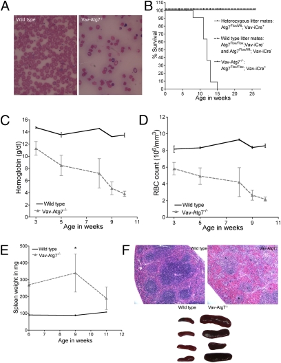
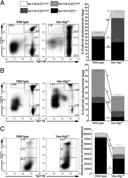
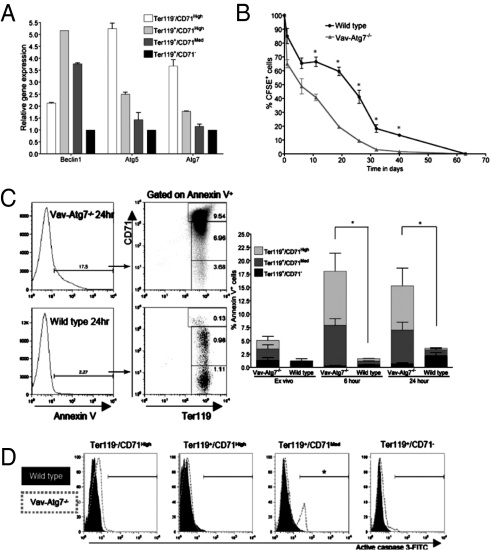
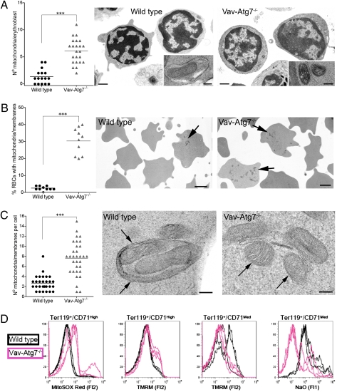
Timely elimination of damaged mitochondria is essential to protect cells from the potential harm of disordered mitochondrial metabolism and release of proapoptotic proteins. In mammalian red blood cells, the expulsion of the nucleus followed by the removal of other organelles, such as mitochondria, are necessary differentiation steps. Mitochondrial sequestration by autophagosomes, followed by delivery to the lysosomal compartment for degradation (mitophagy), is a major mechanism of mitochondrial turnover. Here we show that mice lacking the essential autophagy gene Atg7 in the hematopoietic system develop severe anemia. Atg7(-/-) erythrocytes accumulate damaged mitochondria with altered membrane potential leading to cell death. We find that mitochondrial loss is initiated in the bone marrow at the Ter119(+)/CD71(High) stage. Proteomic analysis of erythrocyte ghosts suggests that in the absence of autophagy other cellular degradation mechanisms are induced. Importantly, neither the removal of endoplasmic reticulum nor ribosomes is affected by the lack of Atg7. Atg7 deficiency also led to severe lymphopenia as a result of mitochondrial damage followed by apoptosis in mature T lymphocytes. Ex vivo short-lived hematopoietic cells such as monocytes and dendritic cells were not affected by the loss of Atg7. In summary, we show that the selective removal of mitochondria by autophagy, but not other organelles, during erythropoeisis is essential and that this is a necessary developmental step in erythroid cells.


Conflict of interest statement

The authors declare no conflict of interest.

Figures

Fig. 1.
Fig. 1.
The absence of Atg7 in the hematopoietic system leads to lethal anemia. (A) Representative blood smears from anemic 11-week-old mice. (B) Survival curves. (C) Hemoglobin levels. (D) Red blood cell counts. (E) Spleen weight in mg (*P = 0.0159). (F) Upper: H&E-stained paraffin-embedded spleen sections. Lower: representative spleen size, 10-week-old mice.
Fig. 2.
Fig. 2.
Altered erythroid developmental stages in Vav-Atg7−/− mice. Distribution of erythroid developmental stages in (A) the spleen of 6-week-old mice (n = 4), (B) the BM of 6-week-old mice (n = 4), and (C) the blood of 9-week-old mice (n = 4). ns: P > 0.05; *P < 0.05. Stages as gated on dot plots, and individual stage frequencies are plotted in bar graphs. One of three representative experiments is shown.
Fig. 3.
Fig. 3.
Atg7−/− erythroid cells have a decreased lifespan and a significantly increased susceptibility to cell death in both blood and spleen. (A) qPCR amplification of Beclin1, Atg5, and Atg7 cDNA in the indicated BM erythroid developmental stages from 8-week-old WT mice. (B) Survival of CFSE-labeled WT and Vav-Atg7−/− red blood cells in WT hosts (*P < 0.05). This experiment was repeated three times with similar results. (C) Annexin V levels in peripheral red blood cells ex vivo, after 6 and 24 h of culture. The percentage of Annexin V+ cells was determined by gating on live cells and is shown in the left panel. Bar heights indicate total percentage of Annexin V+ cells (interaction: *P < 0.05, two-way ANOVA, n = 3). (D) Representative histograms of active caspase 3 levels on spleen erythroid stages from 9-week-old mice, gated as in Fig. 2A (*P = 0.0159, n = 4).
Fig. 4.
Fig. 4.
Atg7−/− erythroid cells accumulate damaged mitochondria. (A) Left: total mitochondrial count/BM erythroblast counted by EM. Right: representative EM. (Scale bars, 1 mm in lower power and 100 nm in inset.) (B) Left: percent of peripheral RBCs containing remnants of mitochondria or other membranes. Right: representative EM. (Scale bars, 1 μm.) Arrows: mitochondrial remnants. (C) Left: number of mitochondria and membranes per cell. Right: representative EM. (Scale bars, 100 nm.) In WT, arrows point at autophagosome; in Atg7−/−, arrows point at mitochondria. (D) Overlays of three WT and three Vav-Atg7−/− BM erythroid developmental stages stained with MitoSOX Red, TMRM, and NaO (gated on the indicated erythroid stages as shown in Fig. 2B) (***P < 0.0001).

References

    1. Kim I, Rodriguez-Enriquez S, Lemasters JJ. Selective degradation of mitochondria by mitophagy. Arch Biochem Biophys. 2007;462:245–253. - PMC - PubMed
    1. Monastyrska I, Klionsky DJ. Autophagy in organelle homeostasis: Peroxisome turnover. Mol Aspects Med. 2006;27:483–494. - PMC - PubMed
    1. Bernales S, McDonald KL, Walter P. Autophagy counterbalances endoplasmic reticulum expansion during the unfolded protein response. PLoS Biol. 2006;4:e423. - PMC - PubMed
    1. Krick R, et al. Piecemeal microautophagy of the nucleus requires the core macroautophagy genes. Mol Biol Cell. 2008;19:4492–4505. - PMC - PubMed
    1. Maiuri MC, Zalckvar E, Kimchi A, Kroemer G. Self-eating and self-killing: Crosstalk between autophagy and apoptosis. Nat Rev Mol Cell Biol. 2007;8:741–752. - PubMed

Publication types

MeSH terms