. 2010 Jan 12;107(2):844-9.
doi: 10.1073/pnas.0909781107. Epub 2009 Dec 18.

Disruption of TAK1 in hepatocytes causes hepatic injury, inflammation, fibrosis, and carcinogenesis

Affiliations

Disruption of TAK1 in hepatocytes causes hepatic injury, inflammation, fibrosis, and carcinogenesis

Sayaka Inokuchi et al. Proc Natl Acad Sci U S A. .

Abstract

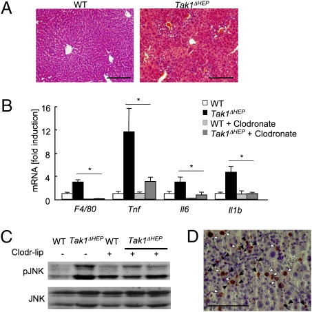
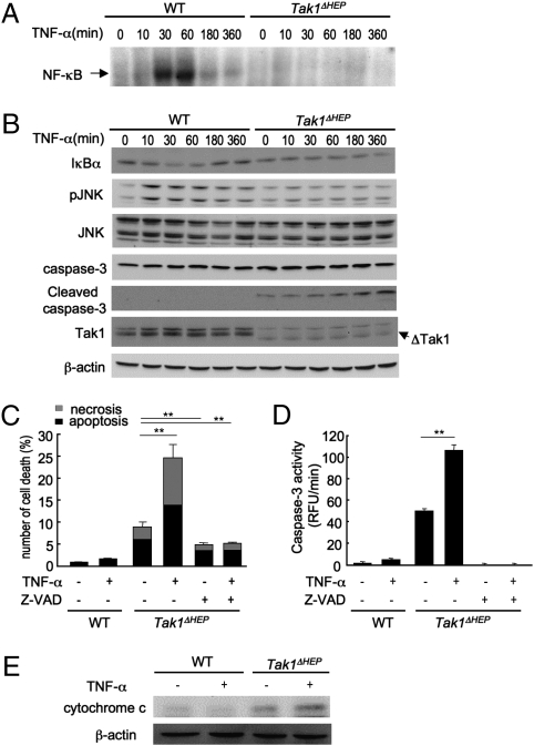
TGF-beta-activated kinase 1 (TAK1) is a MAP3K family member that activates NF-kappaB and JNK via Toll-like receptors and the receptors for IL-1, TNF-alpha, and TGF-beta. Because the TAK1 downstream molecules NF-kappaB and JNK have opposite effects on cell death and carcinogenesis, the role of TAK1 in the liver is unpredictable. To address this issue, we generated hepatocyte-specific Tak1-deficient (Tak1DeltaHEP) mice. The Tak1DeltaHEP mice displayed spontaneous hepatocyte death, compensatory proliferation, inflammatory cell infiltration, and perisinusoidal fibrosis at age 1 month. Older Tak1DeltaHEP mice developed multiple cancer nodules characterized by increased expression of fetal liver genes including alpha-fetoprotein. Cultures of primary hepatocytes deficient in Tak1 exhibited spontaneous cell death that was further increased in response to TNF-alpha. TNF-alpha increased caspase-3 activity but activated neither NF-kappaB nor JNK in Tak1-deficient hepatocytes. Genetic abrogation of TNF receptor type I (TNFRI) in Tak1DeltaHEP mice reduced liver damage, inflammation, and fibrosis compared with unmodified Tak1DeltaHEP mice. In conclusion, hepatocyte-specific deletion of TAK1 in mice resulted in spontaneous hepatocyte death, inflammation, fibrosis, and carcinogenesis that was partially mediated by TNFR signaling, indicating that TAK1 is an essential component for cellular homeostasis in the liver.


Conflict of interest statement

The authors declare no conflict of interest.

Figures

Fig. 1.
Fig. 1.
 Tak1-deficient hepatocytes develop hepatocellular carcinoma. (A and B) TAK1 protein expression in whole liver (A) and primary hepatocytes (B) from WT and Tak1ΔHEP mice was determined by immunoblot analysis. ΔTAK1 indicates a truncated form of TAK1. (C) Livers of 12-month-old WT and Tak1ΔHEP mice. (D and E) Histological findings of 9-month-old WT (D) and Tak1ΔHEP mice (E) by H&E staining. (F) Expression of α-fetoprotein in tumors of Tak1ΔHEP mice assessed by immunohistochemistry. (G and H) Fetal liver gene expression, including Afp (G) and H19, Igf2, Dlk1, and Rex3 (H) in liver tissue from 9-month-old WT (n = 4), and nontumor liver (n = 6) and tumors (n = 6) from Tak1ΔHEP mice, determined by real-time PCR. NT, nontumor liver, T, tumors. Data are reported as mean ± SEM. *P < .05; **P < .01. (Scale bar: 1 cm in C, 20 μm in DF.)
Fig. 2.
Fig. 2.
 Loss of Tak1 in hepatocytes induces spontaneous apoptosis and compensatory regeneration in the liver. (A) Serum ALT levels were measured in WT and Tak1ΔHEP mice at the age 1, 4, and 9 months (n = 10 at the each time point). (BD) WT (n = 10) and Tak1ΔHEP (n = 10) mice at age 1 month were analyzed. (B) Immunoblots of full-length and cleaved caspase-3 and cyclin D1. (C) Apoptotic hepatocytes evaluated by TUNEL staining. (D) Proliferating hepatocytes evaluated by immunohistochemistry for PCNA. Data are reported as mean ± SEM. *P < .05; **P < .01 for Tak1ΔHEP mice versus WT mice. (Scale bar: 20 μm in C and F.)
Fig. 3.
Fig. 3.
 Spontaneous hepatitis in Tak1ΔHEP mice. (AC) WT (n = 10), Tak1ΔHEP (n = 10), and Kupffer cell–depleted Tak1ΔHEP mice (n = 3) were analyzed at age 1 month. Kupffer cells were depleted by liposomal clodronate injection 24 h before death. (A) H&E staining. (B) Hepatic mRNA expression of inflammatory genes including F4/80, Tnf, Il6, and Il1b, determined by real-time PCR. (C) Phospho-JNK and JNK expression in livers from WT, Tak1ΔHEP mice, and Kupffer cell–depleted Tak1ΔHEP mice assessed by immunoblot analysis. (D) Immunohistochemistry for phospho-JNK in Tak1ΔHEP liver. Black and white arrows indicate JNK activation in nonparenchymal cells and hepatocytes, respectively. Data are reported mean ± SEM. *P < .05; **P < .01. (Scale bar: 20 μm in A and D.)
Fig. 4.
Fig. 4.
 Tak1ΔHEP mice develop spontaneous liver fibrosis. (AD) WT and Tak1ΔHEP mice at the age of 1, 4, and 9 months (n = 10 at the each time point) were used for analysis. Fibrillar collagen deposition was determined by Sirius red staining (A) and its quantification (B). Sirius red staining in 4-month-old mice is shown. (C) Expression of αSMA in 1-month-old mice was determined by immunohistochemistry. (D) Hepatic mRNA expression of fibrogenic markers, including Col1A1, Tgfb1, Acta2, and Timp1, was determined by quantitative real-time PCR in 1-month-old WT and Tak1ΔHEP mice. (E) Hepatic mRNA expression of Tgfb1 was determined in Kupffer cell–depleted Tak1ΔHEP mice by real-time PCR. Data are reported as mean ± SEM. *P < .05; **P < .01 for Tak1ΔHEP mice versus WT mice. (Scale bar: 20 μm in A and C.)
Fig. 5.
Fig. 5.
 Deletion of TAK1 in hepatocytes abolishes JNK and NF-κB activation and confers susceptibility to TNF-mediated cell death. (AC) Primary hepatocytes from WT and Tak1ΔHEP mice were incubated with TNF-α (20 ng/mL) for the indicated time. (A) NF-κB binding activity was determined. (B) IκBα, phospho-JNK, JNK, caspase-3, cleaved caspase-3, TAK1, and β-actin were determined by immunoblot analysis. (C) Apoptotic and necrotic cells were determined by Hoechst and PI staining, respectively, and counted 8 h after stimulation with 20 ng/mL TNF-α with or without pretreatment with z-VAD-FMK (50 μM) for 30 min. (D) Caspase-3 activity was measured 6 h after stimulation with 20 ng/mL TNF-α with or without pretreatment with z-VAD-FMK (50 μM) for 30 min. Data are reported as mean ± SEM of triplicate cultures. **P < .01. (E) Cytosolic expression of cytochrome c was determined by immunoblot analysis.
Fig. 6.
Fig. 6.
 Deletion of TNFRI reduces liver injury, apoptosis, and fibrosis in Tak1ΔHEP mice. WT (n = 10), Tak1ΔHEP (n = 10), Tnfr1−/− (n = 5) and Tnfr1−/− Tak1ΔHEP (n = 7) mice age 1 month were used for the analyses. (A) Expression of TAK1 was determined by immunoblot analysis. (B) Serum ALT levels are shown. (C) Immunoblots of full-length and cleaved caspase-3 are shown. (D) mRNA expression of Tnf, Il1b, Ccl2, Il6, and Timp1 was determined by real-time PCR. (E) Expression of phospho-JNK and JNK in liver was assessed by immunoblot analysis. (F) Collagen deposition was determined by quantification of Sirius red staining. Data are reported as mean ± SEM. *P < .05; **P < .01.

Comment in

References

    1. Akira S, Takeda K. Toll-like receptor signalling. Nat Rev Immunol. 2004;4:499–511. - PubMed
    1. Shim JH, et al. TAK1, but not TAB1 or TAB2, plays an essential role in multiple signaling pathways in vivo. Genes Dev. 2005;19:2668–2681. - PMC - PubMed
    1. Ghosh S, Karin M. Missing pieces in the NF-kappaB puzzle. Cell. 2002;109(Suppl):S81–S96. - PubMed
    1. Maeda S, Kamata H, Luo JL, Leffert H, Karin M. IKKbeta couples hepatocyte death to cytokine-driven compensatory proliferation that promotes chemical hepatocarcinogenesis. Cell. 2005;121:977–990. - PubMed
    1. Sakurai T, Maeda S, Chang L, Karin M. Loss of hepatic NF-kappa B activity enhances chemical hepatocarcinogenesis through sustained c-Jun N-terminal kinase 1 activation. Proc Natl Acad Sci USA. 2006;103:10544–10551. - PMC - PubMed

Publication types

Substances

LinkOut - more resources