. 2010 Jan 12;107(2):856-61.
doi: 10.1073/pnas.0911661107. Epub 2009 Dec 22.

VEGFR1-mediated pericyte ablation links VEGF and PlGF to cancer-associated retinopathy

Affiliations

VEGFR1-mediated pericyte ablation links VEGF and PlGF to cancer-associated retinopathy

Renhai Cao et al. Proc Natl Acad Sci U S A. .

Abstract

VEGF coordinates complex regulation of cellular regeneration and interactions between endothelial and perivascular cells; dysfunction of the VEGF signaling system leads to retinopathy. Here, we show that systemic delivery of VEGF and placental growth factor (PlGF) by protein implantation, tumors, and adenoviral vectors ablates pericytes from the mature retinal vasculature through the VEGF receptor 1 (VEGFR1)-mediated signaling pathway, leading to increased vascular leakage. In contrast, we demonstrate VEGF receptor 2 (VEGFR2) is primarily expressed in nonvascular photoreceptors and ganglion cells. Moreover, blockade of VEGFR1 but not VEGFR2 significantly restores pericyte saturation in mature retinal vessels. Our findings link VEGF and PlGF to cancer-associated retinopathy, reveal the molecular mechanisms of VEGFR1 ligand-mediated retinopathy, and define VEGFR1 as an important target of antiangiogenic therapy for treatment of retinopathy.


Conflict of interest statement

The authors declare no conflict of interest.

Figures
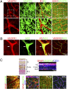
Fig. 1.
Fig. 1.
Differential expression of VEGR1 and VEGFR2 in the retina. (A) VEGFR1 was expressed in the adult retinal vasculature and VEGFR2 was expressed in nonvascular structures including ganglion cells and photoreceptors (green). Retinal blood vessels were revealed by CD31 staining. (B) Colocalization of VEGFR1 (red) and the pericyte marker NG2 (green) in the retinal vasculature. VEGFR2 (red) and NG2 (green) did not show overlapping staining (Far Right). Arrows point to the NG2 and VEGFR1 positive pericytes. (C) GFAP (red) and VEGFR2 (green) exhibit overlapping positive signals in the ganglion layer (GL), the plexiform layer and nuclear layer (PL/NL), and the rods and cones layer (RCL) of retinal tissue. CD31 staining (blue) is also shown.
Fig. 2.
Fig. 2.
Expression and activation of VEGFR1 in isolated pericytes and VSMCs. (A) Isolated primary mouse pericytes (Upper) express the pericyte-specific marker NG2 (green); a subpopulation express αSMA (red). Isolated VSMCs (Lower) express αSMA (red); a subpopulation express NG (green). Merged images are shown at right. (B) Immunocytochemistry indicating that isolated pericytes and VSMCs exhibit positive staining of VEGFR1 (green) and PDGFRβ (red). (C) Reverse transcriptase-PCR analysis of VEGFR1 mRNA expression in isolated mouse pericytes and endothelial cells (SVEC). (D) Western blot analysis demonstrating activation (increased phosphorylation) of Src and Erk and down-regulation (decreased phosphorylation) of Akt in isolated pericytes after VEGF treatment for the indicated time. GAPDH was analyzed as a control. (E) Western blot analysis demonstrating VEGFR1 blockade inhibits VEGF–induced Erk activation (phosphorylation). GAPDH was analyzed as a control.
Fig. 3.
Fig. 3.
Retinal pericyte ablation by local and systemic delivery of VEGF or PlGF. (A) Using the micropocket assay, VEGF or PlGF (160 ng) was implanted into the cornea of each mouse eye. Mouse retinas were isolated on days 5 and 25 after implantation, followed by whole-mount immunofluorescence staining to detect double staining with NG2 (green) and CD31 (red). (B) Untreated retinas (No factor), AdVector-treated retinas, AdVEGF-treated retinas, or retinas treated with neutralizing PDGFR β antibody were double stained with NG2 (green) and CD31 (red). (Scale bars: 50 μm.) (C and D) Quantification of pericyte saturation in retinal blood vessels of mice treated as indicated. Pericytes were detected by NG2 and CD31 double staining and 10 randomized fields were analyzed for quantification per group.
Fig. 4.
Fig. 4.
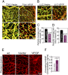
Retinal pericyte ablation and vascular permeability by tumor-derived VEGF. (A) T241-Vector or T241-VEGF tumor cell lines were s.c. injected in the dorsal back of C57BL mice and tumors were allowed to develop. Retinas were harvested when tumor size was approximately 1 cm3 and subjected to whole-mount immunofluorescence staining to detect NG2 (green) and CD31 (red). (Scale bar: 50 μm.) (B) LLC-Vector or LLC-VEGF tumor cell lines were s.c. injected in the backs of C57BL mice. Retinas were harvested when tumor size was approximately 1 cm3 and subjected to whole-mount immunofluorescence staining to detect NG2 (green) and CD31 (red). (Scale bar: 50 μm.) (C) Quantification of pericyte saturation in retinal vessels of mice treated as in A. Approximately 10 randomized fields were quantified per treatment group. (D) Quantification of pericyte saturation in retinal vessels of mice treated as in B. Approximately 10 randomized fields were quantified per treatment group. (E) Vascular permeability of rhodamine-labeled dextran (70 kDa) in the retinas of WT, vector, and VEGF tumor-bearing mice. (Scale bar: 50 μm.) (F) ELISA measurement of plasma VEGF levels in mice treated as in A.
Fig. 5.
Fig. 5.
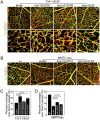
Systemic VEGF-A expression results in retinal pericyte ablation in two mouse models: VEGFR1 blockade results in pericyte recovery. (A) Retinas from T241-VEGF tumor-bearing mice treated with anti-VEGFR1 (MF1), anti-VEGFR2 (DC101), or anti-VEGF (bevacizumab) were double stained with NG2 (green) and CD31 (red). (Scale bar: 50 μm.) (B) Retinas of healthy mice (WT), or MMTV-neu mice harboring spontaneous mammary tumors were treated with buffer, anti-VEGFR1 (MF1) antibody, or anti-VEGFR2 (DC101) antibody and then double stained with NG2 (green) and CD31 (red). (Scale bar: 50 μm.) (C) Quantification of pericyte coverage of vessels from retinas of mice treated as in A. Approximately 10 randomized fields were quantified per treatment group. (D) Quantification of pericyte coverage of vessels from retinas of mice treated as in B. Approximately 10 randomized fields were used for quantification in each group.

References

    1. Alon T, et al. Vascular endothelial growth factor acts as a survival factor for newly formed retinal vessels and has implications for retinopathy of prematurity. Nat Med. 1995;1:1024–1028. - PubMed
    1. Ambati BK, et al. Corneal avascularity is due to soluble VEGF receptor-1. Nature. 2006;443:993–997. - PMC - PubMed
    1. Senger DR, et al. Tumor cells secrete a vascular permeability factor that promotes accumulation of ascites fluid. Science. 1983;219:983–985. - PubMed
    1. Buzney SM, Frank RN, Robison WG., Jr Retinal capillaries: Proliferation of mural cells in vitro. Science. 1975;190:985–986. - PubMed
    1. Hirschi KK, D’Amore PA. Pericytes in the microvasculature. Cardiovasc Res. 1996;32:687–698. - PubMed

Publication types

MeSH terms

Substances