. 2009 Dec 29;106(52):22102-7.
doi: 10.1073/pnas.0902639106. Epub 2009 Dec 22.

Defining the role of syndecan-4 in mechanotransduction using surface-modification approaches

Affiliations

Defining the role of syndecan-4 in mechanotransduction using surface-modification approaches

Robert M Bellin et al. Proc Natl Acad Sci U S A. .

Abstract

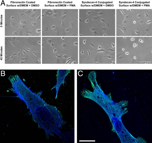
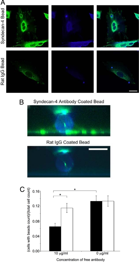
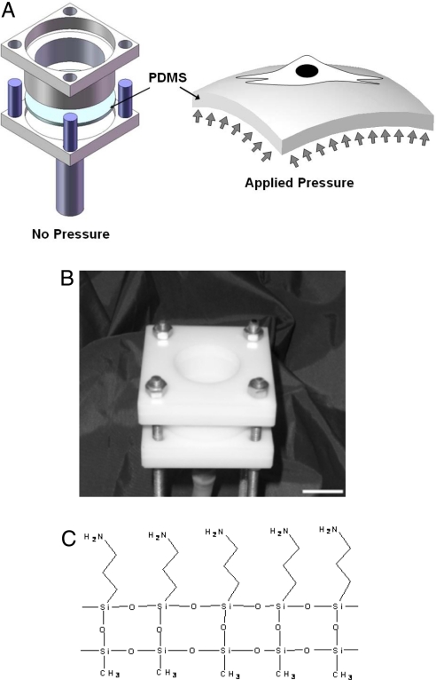
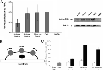
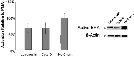
The ability of cells to respond to external mechanical stimulation is a complex and robust process involving a diversity of molecular interactions. Although mechanotransduction has been heavily studied, many questions remain regarding the link between physical stimulation and biochemical response. Of significant interest has been the contribution of the transmembrane proteins involved, and integrins in particular, because of their connectivity to both the extracellular matrix and the cytoskeleton. Here, we demonstrate the existence of a mechanically based initiation molecule, syndecan-4. We first demonstrate the ability of syndecan-4 molecules to support cell attachment and spreading without the direct extracellular binding of integrins. We also examine the distribution of focal adhesion-associated proteins through controlling surface interactions of beads with molecular specificity in binding to living cells. Furthermore, after adhering cells to elastomeric membranes via syndecan-4-specific attachments we mechanically strained the cells via our mechanical stimulation and polymer surface chemical modification approach. We found ERK phosphorylation similar to that shown for mechanotransductive response for integrin-based cell attachments through our elastomeric membrane-based approach and optical magnetic twisting cytometry for syndecan-4. Finally, through the use of cytoskeletal disruption agents, this mechanical signaling was shown to be actin cytoskeleton dependent. We believe that these results will be of interest to a wide range of fields, including mechanotransduction, syndecan biology, and cell-material interactions.


Conflict of interest statement

The authors declare no conflict of interest.

Figures

Fig. 1.
Fig. 1.
NIH 3T3 fibroblasts grown on fibronectin or anti-syndecan-4 antibody-coated glass surfaces. (A) After growing cells on fibronectin or anti-syndecan-4 antibodies, cells were treated with PMA or DMSO as a control. PMA induces the enzyme-driven shedding of syndecans, but not integrins. The rounding and detachment of cells on anti-syndecan-4 antibody surfaces, but not fibronectin surfaces, indicates that integrins are not directly involved in the binding to anti-syndecan-4 antibody-coated surfaces. (Bar: 200 μm.) (B and C) Colocalization of vinculin and actin for NIH 3T3 fibroblasts spread on modified glass surfaces for attachment through transmembrane proteins. NIH 3T3 cells on surfaces modified with fibronectin (B) and anti-syndecan-4 antibodies (C) were immunofluorescently labeled for vinculin (green) and phalloidin for actin (blue). Note that cells attached exclusively through syndecan-4 recruit vinculin to sites of focal contact. (Bar: 25 μm.)
Fig. 2.
Fig. 2.
Localization of structural proteins with syndecan-4 through bead binding. (A) NIH 3T3 cells were grown on glass and incubated for 45 min at 37 °C with syndecan-4 antibody or rat IgG-conjugated beads. Cells were fixed and labeled for vinculin (green) and actin (blue). Note the vinculin halo structure and increased actin labeling surrounding syndecan-4 antibody beads and the lack of such labeling surrounding rat IgG bead. (Bar: 25 μm.) (B) An 8-μm side view of bead attachments from A created by reconstruction of 100 × 0.08-μm confocal slices. (Bar: 5 μm.) (C) Free anti-syndecan-4 antibodies (filled bars) inhibited anti-syndecan-4 conjugated beads from binding to NIH 3T3 cells, whereas free rat IgG antibodies (empty bars) did not (*, P < 0.05).
Fig. 3.
Fig. 3.
Mechanical stimulation of NIH 3T3 fibroblasts used to examine syndecan-4-based mechanotransduction. (A and B) Schematics (A) and image (B) of the PreCS device for mechanical stimulation of individual cells on their basal domains through stretching a PDMS membrane. (Scale bar: 2 cm.) (C) Surface modification method for attaching living cells to the elastomeric membranes with molecular specificity. The molecular attachment was accomplished by using an AMEO modification of PDMS, which then enabled the direct attachment of anti-syndecan-4 antibodies to the polymer surface.
Fig. 4.
Fig. 4.
Activation of ERK under mechanical stimulation through syndecan-4. (A) Western blot and densitometry analysis for mechanical strain that was applied to the NIH 3T3 cells attached through anti-syndecan-4 antibodies for 0, 5, and 30 min. PMA-treated cells are a positive control for ERK phosphorylations, and the resulting level of relative activation was defined as 100% for comparison between experimental trials. DMSO-treated cells were a negative control for chemical activation of ERK phosphorylation. The results for a 0-min strain were significantly different from the 5-min strain (P < 0.025) and the 30-min strain (P < 0.025) based on t tests of log10-transformed data with a Bonforroni correction (α = 0.05/2) to treat for repeated testing. Each bar represents the average of at least three independent experiments. (B and C) Using an OMTC device (B) we obtained ERK activation measurements (C) with ELISA-based analysis of lysates of NIH 3T3 cells subjected to strain by twisting coated beads at 1 Hz for 30 min. The resulting ELISA data were normalized based on lysate protein concentration, and then the resulting values were scaled based on defining the twist treatment of fibronectin beads as 100% activation. The resulting ELISA T tests of selected comparisons among the resulting data show that the twist treatment of anti-syndecan-4-coated beads was significantly different with the no-bead sample (P < 0.0167), the no-twist anti-syndecan-4-coated bead sample (P < 0.0167), and the twist AcLDL-coated bead sample (P < 0.0167), using a Bonferroni correction (α = 0.05/3) to treat for repeated testing. Each bar represents the average of three independent experiments.
Fig. 5.
Fig. 5.
Effects of disruption of the actin cytoskeleton with mechanical stimulation through syndecan-4. Shown are Western blot and densitometry analysis of ERK phosphorylation with latrunculin-B and cytochalasin-D treatment under 30 min of strain. The level of ERK phosphorylation in cells that were not chemically treated but mechanically stimulated was statistically higher than both the latrunculin-treated cells (P < 0.025) and the cytochalasin-D treated cells (P < 0.025) based on t tests with a Bonforroni correction (α = 0.05/2) to treat for repeated testing. Each bar represents the average of at least four independent experiments.

Similar articles

Cited by

References

    1. Hahn C, Schwartz MA. Mechanotransduction in vascular physiology and atherogenesis. Nat Rev Mol Cell Biol. 2009;10:53–62. - PMC - PubMed
    1. Wang Y, et al. Visualizing the mechanical activation of Src. Nature. 2005;434:1040–1045. - PubMed
    1. Engler A, Sen S, Sweeney HL, Discher DE. Matrix elasticity directs stem cell lineage specification. Cell. 2006;126:677–689. - PubMed
    1. Ferrer I, et al. Active, phosphorylation-dependent mitogen-activated protein kinase (MAPK/ERK), stress-activated protein kinase/c-Jun N-terminal kinase (SAPK/JNK), and p38 kinase expression in Parkinson's disease and dementia with Lewy bodies. J Neural Transm. 2001;108:1383–1396. - PubMed
    1. Li C, Hu Y, Mayr M, Xu Q. Cyclic strain stress-induced mitogen-activated protein kinase (MAPK) phosphatase 1 expression in vascular smooth muscle cells is regulated by Ras/Rac-MAPK pathways. J Biol Chem. 1999;274:25273–25280. - PubMed

Publication types

MeSH terms

LinkOut - more resources