. 2010 Feb 1;24(3):241-55.
doi: 10.1101/gad.1874010. Epub 2010 Jan 15.

IL-4 induces cathepsin protease activity in tumor-associated macrophages to promote cancer growth and invasion

Affiliations

IL-4 induces cathepsin protease activity in tumor-associated macrophages to promote cancer growth and invasion

Vasilena Gocheva et al. Genes Dev. .

Abstract

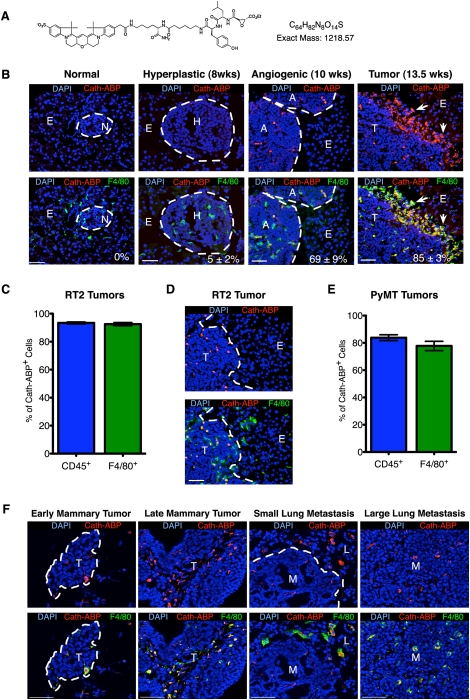
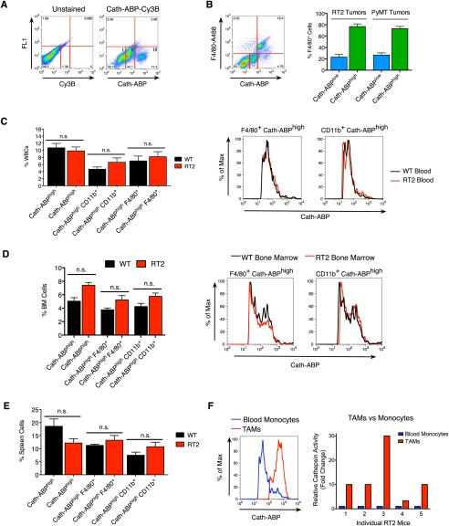
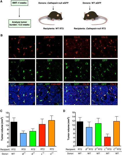
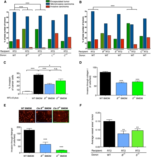
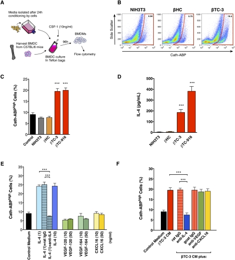
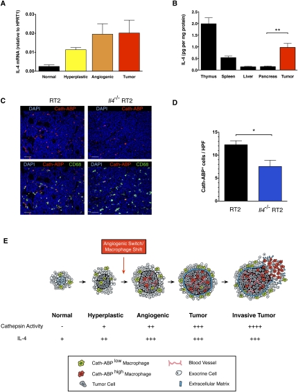
Innate immune cells can constitute a substantial proportion of the cells within the tumor microenvironment and have been associated with tumor malignancy in patients and animal models of cancer; however, the mechanisms by which they modulate cancer progression are incompletely understood. Here, we show that high levels of cathepsin protease activity are induced in the majority of macrophages in the microenvironment of pancreatic islet cancers, mammary tumors, and lung metastases during malignant progression. We further show that tumor-associated macrophage (TAM)-supplied cathepsins B and S are critical for promoting pancreatic tumor growth, angiogenesis, and invasion in vivo, and markedly enhance the invasiveness of cancer cells in culture. Finally, we demonstrate that interleukin-4 (IL-4) is responsible for inducing cathepsin activity in macrophages in vitro and in vivo. Together, these data establish IL-4 as an important regulator, and cathepsin proteases as critical mediators, of the cancer-promoting functions of TAMs.


Figures

Figure 1.
Figure 1.
Increase in cathepsin activity in TAMs during pancreatic islet and mammary cancer development. (A) Chemical structure of Cath-ABP. (B) Cathepsins are highly activated in infiltrating macrophages during tumor progression at the angiogenic islet and tumor stages of RT2 tumorigenesis. Pancreatic tissue from mice injected with the Cath-ABP was stained with an α-F4/80 antibody. Groups of four or more mice were analyzed per stage and three or more lesions per mouse. Normal (N) Tag+ islets were analyzed at 4–7 wk, hyperplastic (H) islets at 8 wk, angiogenic (A) islets at 10 wk, and tumors (T) at 13.5 wk of age. The percentage of F4/80+ cells that were Cath-ABP+ was determined by image analysis and is indicated in the image for each stage. Macrophages present in the normal exocrine (E) pancreas were omitted from the analysis. Arrows represent the invasive tumor front. (C) Graph showing the percent of Cath-ABP+ cells in RT2 tumors that colocalize with F4/80 or CD45, as determined by image analysis using TissueGnostics software (n = 18 tumors). Data are represented as the mean ± SEM. (D) Representative image of a tumor (T) from a 13.5-wk-old RT2 mouse showing macrophages inside the lesion with high levels of cathepsin activity as compared with those in the adjacent normal exocrine (E) pancreas. (E) Graph showing the percent of Cath-ABP+ cells in PyMT tumors that colocalize with CD45 (n = 10 tumors from four independent mice) or F4/80 (n = 9 tumors from four independent mice), analyzed as in C. (F) Cathepsins are highly activated in TAMs during mammary tumor (T) development and lung metastasis (M) in PyMT mice. Early mammary tumors were obtained from 9-wk-old mice, late tumors were obtained from 14-wk-old mice, and small and large lung metastases were obtained from 14-wk old mice, which have variable lung metastatic burdens. The adjacent normal lung (L) parenchyma is shown for comparison. Bars, 50 μm.
Figure 2.
Figure 2.
Macrophages infiltrating the local tumor microenvironment of pancreatic islet tumors have high levels of cathepsin activity. (A) Flow cytometry was used to label whole-tumor suspensions from RT2 mice ex vivo with the Cath-ABP. Subsequent analysis revealed three different levels of cathepsin activation in whole tumors: negative/low (I), medium (II), and high (III) (n = 13 mice). (B) The majority of macrophages present inside RT2 and PyMT tumors have high levels of cathepsin activity. Whole-tumor suspensions were labeled with the Cath-ABP and α-F4/80, and the proportion of macrophages with high and low levels of cathepsin activity was quantified by flow cytometry. A representative plot from a RT2 sample is shown on the left (RT2 mice, n = 7; PyMT mice, n = 9). (C) Cathepsin activity is not elevated in circulating monocytes from RT2 tumor-bearing mice compared with wild-type (WT) mice. WBCs isolated from wild-type and RT2 mice were labeled ex vivo with the Cath-ABP, α-F4/80, and α-CD11b antibodies. The number of Cath-ABPhigh cells in the blood (graph) and the level of activation (flow cytometry plots) were determined; (n.s.) not significant (n > 5 mice). (D) Cathepsin proteases are not significantly activated in the BM of RT2 mice when compared with wild-type littermates. BM cells from wild-type and RT2 mice were stained and analyzed as in C. (E) Cathepsin proteases are not significantly activated in the spleen of RT2 mice when compared with wild-type littermates. Spleen cells from wild-type and RT2 mice were stained as in C. (F) The level of cathepsin activity is enhanced in TAMs compared with blood monocytes of RT2 mice. Cathepsin activation was determined by labeling blood and tumor suspensions from individual RT2 mice with the Cath-ABP and α-F4/80. The mean fluorescence intensity of cathepsin activity was determined for TAMs and blood monocytes from each mouse, and the relative cathepsin activity in TAMs versus blood monocytes is shown in the graph for five individual animals. Cathepsin activity was 12.7-fold higher on average in TAMs compared with monocytes; P = 0.028.
Figure 3.
Figure 3.
Removal of BM-derived Cts B and S significantly impairs RT2 tumor progression. (A) Schematic of the BMT experimental design. (B) The vast majority of BMDCs that infiltrate tumors of RT2 mice transplanted with GFP+ wild-type (WT) BM differentiate into F4/80+ macrophages, which supply cathepsin activity and express Cts B and S as indicated by immunofluorescence analysis. Bar, 50 μm. (C) Tumor burden was decreased in RT2 mice transplanted with Cts B- or S-null BM. The total number of BMT recipient mice analyzed were as follows: wild type (WT), 24; B−/−, 20 (P < 0.05); S−/−, 24; L−/−, 15; C−/−, 18. (D) Supplying wild-type BM to individual Cts B- or S-null, but not L-null, RT2 mice restored the tumor burden to wild-type RT2 levels. The following number of BMT recipient mice were analyzed: wild-type (WT) RT2, 24; B−/− RT2, 12; S−/− RT2, 13; L−/− RT2, 3; C−/− RT2, 15.
Figure 4.
Figure 4.
Removal of macrophage-derived Cts B or S impairs cancer cell invasion and angiogenesis. (A) H&E staining was used to grade tumors from BM-transplanted RT2 mice. Graph showing the relative proportions of encapsulated, microinvasive, and invasive carcinomas in RT2 mice that received wild-type (WT) BM compared with those that received Cts-null BM. Deletion of Cts B or S in the BM resulted in a substantial decrease in tumor invasiveness ([***] P ≤ 0.0001 compared with wild type), whereas removal of Cts C or L from the BM had no significant effect. (B) Invasion grading and analysis was performed on Cts-null RT2 mice transplanted with wild-type BM. Transfer of wild-type BM into Cts B- or S-null RT2 recipients restored tumor invasiveness to wild-type RT2 levels, but had no effect on the reduced tumor invasion observed in Cts L-null RT2 mice; (***) P < 0.0001 compared with wild type. (C) Removal of Cts B or S from macrophages reduced macrophage-induced invasion of breast cancer cell line MTLn3. The ability of BMDMs isolated from wild-type, Cts B−/− or S−/− mice to promote invasion of cancer cells into an overlying collagen I matrix was determined. Cancer cells invading >15 μm were scored as invasive in three independent experiments performed in triplicate; (*) P < 0.05; (***) P < 0.0001 compared with wild type. (D) Deletion of Cts B or S in BMDMs reduced macrophage invasion through collagen I by 46% and 40% decrease, respectively, compared with wild type. BMDMs isolated from wild-type, Cts B−/−, or S−/− mice were allowed to invade through collagen I-coated inserts. (***) P < 0.0001 compared with wild type. (E) Deletion of Cts B or S in macrophages reduced their invasion through Matrigel by 63% and 89% decrease, respectively, compared with wild type. BMDMs isolated from wild-type, Cts B−/−, or S−/− mice were allowed to invade through Matrigel-coated inserts. (***) P < 0.0001 compared with wild type. Bar, 100 μm. (F) Transplantation of Cts B- or S-null BM reduces tumor vascularization in the recipient tumors. The tumor area covered by CD31+ cells was analyzed with MetaMorph software. (**) P < 0.01 compared with wild type; n = 5 mice in each group. (C–F) Data are represented as mean and SEM.
Figure 5.
Figure 5.
IL-4 induces cathepsin activity in macrophages. (A) Experimental scheme of the cell-based assay used to investigate the induction of cathepsin activity in BMDMs. (B) Representative flow cytometry plots using the Cath-ABP to assess cathepsin activity in BMDMs incubated with CM prepared from three different cell lines—3T3 fibroblasts, βHC hyperplastic cells, and βTC-3 tumor cells—demonstrating an increased proportion of Cath-ABPhigh BMDMs following incubation with βTC-3 CM. (C) Incubation of BMDCs with βTC-3 or βTC-916 CM resulted in a significant induction of cathepsin activity compared with either βHC or 3T3 CM. (D) The IL-4 levels in CM were measured by ELISA, revealing significantly higher levels of IL-4 in βTC CM than either βHC or 3T3 CM. (E) BMDCs were incubated with various recombinant proteins at indicated concentrations for 96 h, and cathepsin activity was then determined by flow cytometry. Incubation with IL-4 significantly up-regulated cathepsin activity in BMDMs, while addition of a neutralizing antibody against IL-4, but not the isotype control IgG, abolished the effect of IL-4. Incubation with VEGF-120, VEGF-164, and CXCL16 had no effect. (F) BMDCs were incubated with βTC-3 CM plus neutralizing antibodies against IL-4, VEGF, or CXCL16 or the relevant isotype control IgG. The inductive effect of βTC-3 CM on cathepsin activity in BMDMs was completely abrogated by addition of the IL-4 antibody, but not by antibodies to VEGF or CXCL16. (C–F) Data are represented as mean and SEM of three independent experiments performed in duplicate; (***) P < 0.0001.
Figure 6.
Figure 6.
IL-4 levels progressively increase during tumor development and deletion of Il4 reduces cathepsin activity levels in vivo. (A) The levels of IL-4 expression increase during tumor progression in RT2 mice. RNA amplified from pools of lesions at the different stages (n = 3–5) was analyzed using real-time quantitative RT–PCR. (B) IL-4 levels in lysates prepared from RT2 tumors or normal tissues were determined by ELISA, revealing a significant increase in IL-4 produced by tumors compared with normal pancreas; (**) P < 0.01. Data are normalized to the total protein concentration. Thymus and spleen tissues, where there are abundant T cells, were used as positive controls for IL-4 (n = 6–12 mice per group). (C) Representative images showing a decrease in the number of Cath-ABP+ cells within the tumors of Il4−/− RT2 mice as compared with Il4+/+ RT2 controls. The infiltrating macrophages are costained with CD68. Bar, 50 μm. (D) The number of Cath-ABP+ cells within the tumors of RT2 and Il4−/− RT2 mice was quantified by image analysis showing a 38.6% decrease; (*) P < 0.05. Data represent mean and SEM per 40× high-power field (HPF) (n = 6 mice per group). (E) Model depicting the induction of cathepsin activity (as determined by Cath-ABP labeling) in infiltrating macrophages. This transition occurs concomitant with angiogenic switching in developing tumors, in response to increased expression of IL-4. Cath-ABP+ macrophages are also found at the invasive edges of tumors, where they enhance cancer cell invasion by degradation of ECM proteins.

References

    1. Buck MR, Karustis DG, Day NA, Honn KV, Sloane BF. Degradation of extracellular-matrix proteins by human cathepsin B from normal and tumour tissues. Biochem J. 1992;282:273–278. - PMC - PubMed
    1. DeNardo DG, Barreto JB, Andreu P, Vasquez L, Tawfik D, Kolhatkar N, Coussens LM. CD4+ T cells regulate pulmonary metastasis of mammary carcinomas by enhancing protumor properties of macrophages. Cancer Cell. 2009;16:91–102. - PMC - PubMed
    1. Efrat S, Linde S, Kofod H, Spector D, Delannoy M, Grant S, Hanahan D, Baekkeskov S. β-Cell lines derived from transgenic mice expressing a hybrid insulin gene–oncogene. Proc Natl Acad Sci. 1988;85:9037–9041. - PMC - PubMed
    1. Gocheva V, Zeng W, Ke D, Klimstra D, Reinheckel T, Peters C, Hanahan D, Joyce JA. Distinct roles for cysteine cathepsin genes in multistage tumorigenesis. Genes & Dev. 2006;20:543–556. - PMC - PubMed
    1. Goswami S, Sahai E, Wyckoff JB, Cammer M, Cox D, Pixley FJ, Stanley ER, Segall JE, Condeelis JS. Macrophages promote the invasion of breast carcinoma cells via a colony-stimulating factor-1/epidermal growth factor paracrine loop. Cancer Res. 2005;65:5278–5283. - PubMed

Publication types

LinkOut - more resources