. 2010 Jan 15;24(2):171-82.
doi: 10.1101/gad.552910.

Cooperative interaction between retinoic acid receptor-alpha and estrogen receptor in breast cancer

Affiliations

Cooperative interaction between retinoic acid receptor-alpha and estrogen receptor in breast cancer

Caryn S Ross-Innes et al. Genes Dev. .

Abstract

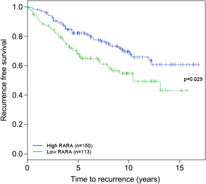
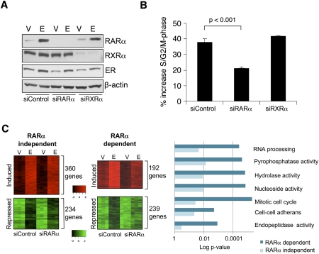
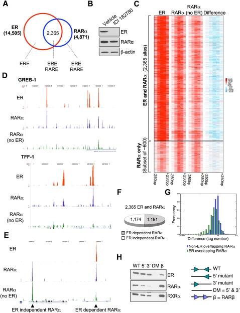
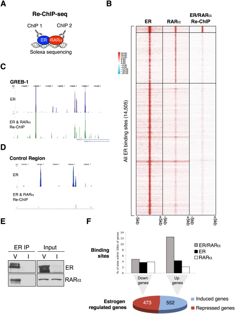
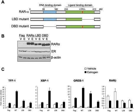
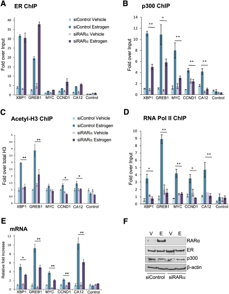
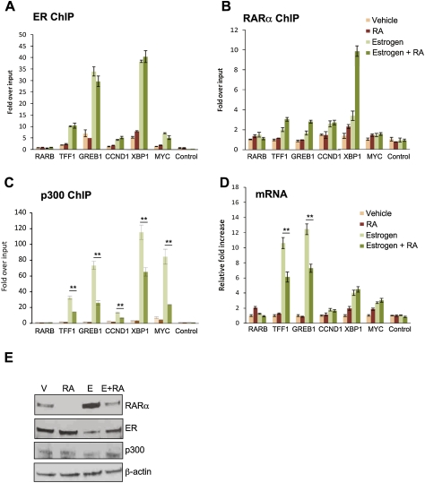
Retinoic acid receptor-alpha (RAR alpha) is a known estrogen target gene in breast cancer cells. The consequence of RAR alpha induction by estrogen was previously unknown. We now show that RAR alpha is required for efficient estrogen receptor-alpha (ER)-mediated transcription and cell proliferation. RAR alpha can interact with ER-binding sites, but this occurs in an ER-dependent manner, providing a novel role for RAR alpha that is independent of its classic role. We show, on a genome-wide scale, that RAR alpha and ER can co-occupy regulatory regions together within the chromatin. This transcriptionally active co-occupancy and dependency occurs when exposed to the predominant breast cancer hormone, estrogen--an interaction that is promoted by the estrogen-ER induction of RAR alpha. These findings implicate RAR alpha as an essential component of the ER complex, potentially by maintaining ER-cofactor interactions, and suggest that different nuclear receptors can cooperate for effective transcriptional activity in breast cancer cells.


Figures

Figure 1.
Figure 1.
RARα predicts the outcome in breast cancer patients. RARα expression levels in 263 breast cancer patients, all of whom received endocrine therapy, were used to predict positive clinical outcome (recurrence-free survival).
Figure 2.
Figure 2.
RARα is required for estrogen-mediated transcription and proliferation. (A) Western blot analysis of nuclear extracts of MCF-7 cells transfected with control siRNA or siRNA to RARα or RXRα and treated with vehicle (V) or estrogen (E). (B) Control siRNA or siRNA to RARα or RXRα was transfected into hormone-deprived cells, followed by estrogen treatment and assessment of percentage of S/G2/M phase. The data are the average of three independent replicates, and the error bars represent standard deviation (SD). (C) siControl- or siRARα-transfected cells were treated with vehicle or estrogen, and gene expression profiling was performed. Examples of estrogen-induced or estrogen-repressed genes are shown based on their dependency on RARα. GO category enrichments of the differentially estrogen-regulated genes (those dependent on RARα vs. those independent) are shown.
Figure 3.
Figure 3.
RARα binds to ER-binding regions in chromatin. (A) ER- and RARα-binding events were mapped in asynchronous MCF-7 cells. The ERE and RARE motifs that were enriched within each of the categories are shown. (B) Cells were treated with vehicle or ICI 182780, an ER down-regulator, for 3 h. Total protein was analyzed using Western blot analysis. (C) Clustering of ER and RARα binding ±250 bp. Signal based on tag count within all ER-binding events and corresponding RARα binding in full media or ICI 182780-treated cells. The difference in tag number within RARα-binding events between full media and ICI 182780 treatments is shown. As a control, ∼600 RARα-binding events (but not ER-binding events) are shown, and these are not influenced by the presence of ICI 182780. (D) Examples of ER- and RARα-binding events adjacent to the TFF-1 and GREB-1 genes. In both cases, the down-regulation of ER results in significant decreases in RARα binding. (E) A control region showing two RARα-binding events: one that does not overlap with ER binding and is ER-independent, and the adjacent site that is ER-dependent. (F) Graph representing the percentage of RARα-binding events that require ER. (G) Frequency histogram plotting the difference values (RARα in ICI 182780-treated cells minus RARα signal in full media) comparing RARα-binding events that overlap with ER sites versus non-ER-overlapping RARα-binding events. (H) An in vitro oligonucleotide pull-down was performed using a biotinylated 38-bp double-stranded oligonucleotide of an experimentally verified ER-binding region containing a perfect ERE. Various mutants lacking the 5′ half-site, the 3′ half-site, or both half-sites were included. Also included was a positive RARα/RXRα-binding site (RARβ promoter). Asynchronous MCF-7 cell lysate was collected and incubated with the various oligonucleotides, and purified protein was subjected to Western blot analysis.
Figure 4.
Figure 4.
Genome-wide Re-ChIP-seq shows that ER and RARα co-occupy the chromatin together. (A) A schematic showing the Re-ChIP-seq protocol, where ER ChIP is performed, followed by RARα ChIP and Solexa sequencing. (B) Clustered binding signal from all ER-binding events (±5 kb) is shown. The corresponding RARα-binding events as well as the ER/RARα Re-ChIP binding signal are shown. The signal from the ER/RARα Re-ChIP sites are after the ER/IgG signal has been subtracted. (C) An example of a region around the GREB-1 gene showing the ER/RARα Re-ChIP signal. (D) A control region showing a strong ER-binding events but no ER/RARα Re-ChIP signal. (E) Coimmunoprecipitation showing direct ER and RARα interactions in asynchronous cells treated with vehicle (V). As a control, cells were treated with ICI 182780 (I). Also shown are total input levels. (F) The ER/RARα Re-ChIP binding events, the ER-binding events (not Re-ChIP sites), and the RARα-binding events (not Re-ChIP sites) were assessed relative to estrogen-induced or estrogen-repressed genes. The percentage of binding sites within 50 kb of the transcription start sites of the estrogen-regulated genes are shown.
Figure 5.
Figure 5.
Expression of RARα can potentiate estrogen–ER-mediated transcription. (A) Schematic representation of the RARα mutants generated. (B) Western blot analysis of nuclear fraction of transfected cells after vehicle (V) or estrogen (E) treatment. (C) Hormone-depleted cells were transfected with control (Flag), RARα (WT), or the LBD and DBD mutants of RARα, and cells were subsequently treated with vehicle or estrogen. Real-time RT–PCR was conducted of TFF-1, XBP-1, and GREB-1 (all estrogen-regulated genes). RARβ (a RARα target gene) was included as a control gene.
Figure 6.
Figure 6.
RARα is required for effective coactivator loading. MCF-7 cells were hormone-depleted and transfected with control siRNA or siRNA targeting RARα. ChIP of ER (A), p300 (B), or acetylated-H3 (C) was performed after 6 h of vehicle (V) or estrogen (E) treatment, followed by real-time PCR of a number of ER-binding regions. The acetylated-H3 ChIP was first normalized to total H3, then to Input. (D) Following transfection, RNA PolII ChIP was performed, and real-time PCR of the promoter regions of the estrogen-regulated genes was performed. (E) Changes in mRNA levels of the genes were assessed following silencing of RARα. (F) After silencing of control or RARα, total protein levels of ER, p300, and RARα were assessed. (*) P < 0.05; (**) P < 0.01. The data are representative of triplicate experiments, ±SD.
Figure 7.
Figure 7.
RARα ligands can inhibit estrogen–ER-mediated transcriptional activity. Hormone-depleted cells were treated with vehicle, estrogen, RA (ATRA), or estrogen + RA for 6 h, and ChIP was performed for ER (A), RARα (B), and p300 (C). Real-time PCR of a number of ER-binding regions was assessed. (D) Changes in mRNA levels of several regulated genes. (E) Western blot of total protein levels in treated cells. The data are the average of three independent experiments, ±SD. (*) P < 0.05; (**) P < 0.01.

References

    1. Backes C, Keller A, Kuentzer J, Kneissl B, Comtesse N, Elnakady YA, Muller R, Meese E, Lenhof HP. GeneTrail—Advanced gene set enrichment analysis. Nucleic Acids Res. 2007;35:W186–W192. doi: 10.1093/nar/gkm323. - DOI - PMC - PubMed
    1. Balmer JE, Blomhoff R. A robust characterization of retinoic acid response elements based on a comparison of sites in three species. J Steroid Biochem Mol Biol. 2005;96:347–354. - PubMed
    1. Bastien J, Rochette-Egly C. Nuclear retinoid receptors and the transcription of retinoid-target genes. Gene. 2004;328:1–16. - PubMed
    1. Benjamini Y, Hochberg Y. Controlling the false discovery rate: A practical and powerful approach to multiple testing. J R Stat Soc Ser B Methodol. 1995;57:289–300.
    1. Borrow J, Goddard AD, Sheer D, Solomon E. Molecular analysis of acute promyelocytic leukemia breakpoint cluster region on chromosome 17. Science. 1990;249:1577–1580. - PubMed

Publication types