. 2010 Mar 19;285(12):8665-74.
doi: 10.1074/jbc.M109.058685. Epub 2010 Jan 15.

Agonist-regulated cleavage of the extracellular domain of parathyroid hormone receptor type 1

Affiliations

Agonist-regulated cleavage of the extracellular domain of parathyroid hormone receptor type 1

Christoph Klenk et al. J Biol Chem. .

Abstract

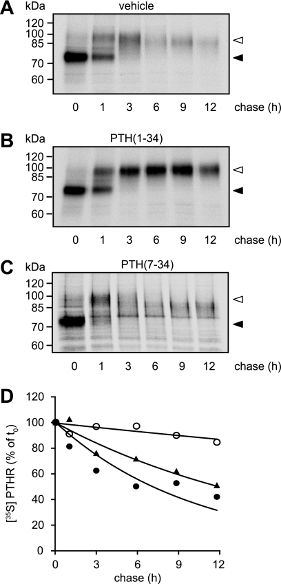
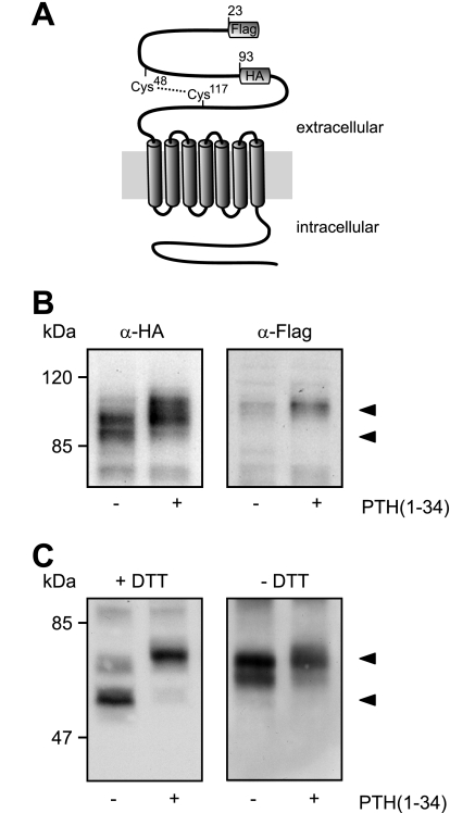
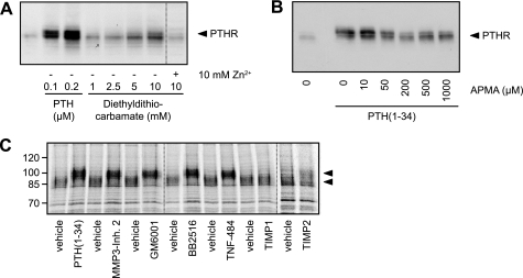
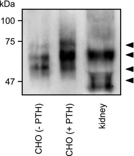
The receptor for parathyroid hormone (PTHR) is a main regulator of calcium homeostasis and bone maintenance. As a member of class B of G protein-coupled receptors, it harbors a large extracellular domain, which is required for ligand binding. Here, we demonstrate that the PTHR extracellular domain is cleaved by a protease belonging to the family of extracellular metalloproteinases. We show that the cleavage takes place in a region of the extracellular domain that belongs to an unstructured loop connecting the ligand-binding parts and that the N-terminal 10-kDa fragment is connected to the receptor core by a disulfide bond. Cleaved receptor revealed reduced protein stability compared with noncleaved receptor, suggesting degradation of the whole receptor. In the presence of the agonistic peptides PTH(1-34), PTH(1-14), or PTH(1-31), the processing of the PTHR extracellular domain was inhibited, and receptor protein levels were stabilized. A processed form of the PTHR was also detected in human kidney. These findings suggest a new model of PTHR processing and regulation of its stability.


Figures

FIGURE 1.
FIGURE 1.
Ligand-induced internalization of the PTHR. A, CHO cells stably expressing HA-PTHR were treated with 100 nm PTH(1–34), 1 μm PTH(1–31), 3 μm PTH(1–14), or 1 μm PTH(7–34) for 30 min. Subsequently, the cells were fixed, permeabilized, and stained with mouse anti-HA antibody followed by a Cy2-labeled anti-mouse antibody. HA-PTHR was visualized by confocal microscopy. B, CHO cells stably expressing HA-PTHR were left untreated (control) or were treated with 100 nm PTH(1–34) or 1 μm PTH(7–34) for 30 min, 3 h, or 6 h. The cells were fixed and either permeabilized with MeOH or left unpermeabilized. White scale bars represent 5 μm. PTHR was visualized as described above.
FIGURE 2.
FIGURE 2.
Prolonged agonist stimulation results in reduced electrophoretic mobility of the PTHR. A and B, CHO cells stably expressing HA-PTHR were treated with 100 nm PTH(1–34), 1 μm PTH(1–31), 3 μm PTH(1–14), or 1 μm PTH(7–34) for the indicated times. Subsequently, the cells were lysed, and the proteins were separated by SDS-PAGE and blotted to polyvinylidene difluoride membranes. PTHR was detected in Western blots with an anti-HA antibody. C, ROS 17/2.8 cells stably expressing HA-PTHR were treated with 100 nm PTH(1–34) for the indicated times. The cells were processed as described above. D, CHO cells stably expressing HA-PTHR or wild type PTHR were treated with 100 nm PTH(1–34) as indicated for 12 h and processed as above. PTHR was detected in Western blot using antibodies detecting an epitope at the PTHR C terminus. The positions of molecular weight standards are marked on the left.
FIGURE 3.
FIGURE 3.
Glycosylation does not contribute to the PTH-induced changes in molecular mass of the PTHR. CHO cells stably expressing HA-PTHR were treated with 100 nm PTH(1–34) for 12 h as indicated. The cells were lysed, and PTHR was precipitated using anti-HA affinity beads. Precipitated proteins were incubated with 50 units/ml of neuraminidase (N), 250 units/ml of Endo H (E), or 5 units/ml of PNGase F (P) for 16 h. Enzyme reactions were stopped by the addition of SDS sample buffer, and proteins were separated by SDS-PAGE. HA-PTHR was detected in Western blot analysis. The empty arrowhead indicates a fully glycosylated receptor form. The closed arrowheads indicate the different receptor forms after PNGase treatment. The positions of molecular weight standards are marked on the left.
FIGURE 4.
FIGURE 4.
PTH stabilizes the high molecular mass form of the PTHR. A–C, CHO cells stably expressing HA-PTHR were pulse-labeled with [35S]methionine/cysteine-containing medium (150 μCi/ml) and then chased for the indicated times with medium containing nonradioactive methionine and cysteine in the absence (A) or presence of 100 nm PTH(1–34) (B) or 1 μm PTH(7–34) (C). HA-PTHR was precipitated from cell lysates using HA antibody. The samples were analyzed by SDS-PAGE and fluorography of the dried gels with a Bio-Rad phosphorimager. The closed arrowheads indicate newly synthesized PTHR; the open arrowheads indicate fully mature PTHR. D, decay of radiolabeled PTHR was quantified using a monoexponential decay function. ▴, untreated; ○, PTH(1–34); ●, PTH(7–34). The means of three independent experiments are shown.
FIGURE 5.
FIGURE 5.
Modification of the PTHR is restricted to the extracellular domain. A, schematic representation of the PTHR. A FLAG epitope was inserted after the signal sequence (amino acids 22 and 23), and an HA epitope was inserted replacing amino acids 93–101. A disulfide bridge between Cys48 and Cys117 stabilizing the extracellular domain is depicted by a dotted line. B, CHO cells stably expressing the FLAG-HA-PTHR were stimulated with PTH(1–34) for 12 h. The cells were lysed, and the samples were subjected to SDS-PAGE and subsequent Western blot analysis. PTHR was detected using an anti-HA antibody (left panel) or an anti-FLAG-M2 antibody (right panel). C, CHO cells stably expressing the HA-PTHR were stimulated with PTH(1–34) for 12 h. To gain better resolution of the proteins, the cells were treated 12 h prior to and throughout the experiment with 5 μg/ml tunicamycin to block N-glycosylation. The cells were lysed in SDS-buffer in the presence (left panel) or absence (right panel) of 100 mm dithiothreitol (DTT). The lysates were subjected to SDS-PAGE and subsequent Western blot analysis. PTHR was detected using an anti-HA antibody. The PTHR forms are indicated by arrowheads.
FIGURE 6.
FIGURE 6.
PTHR is cleaved by an extracellular, zinc-dependent protease. A, CHO cells stably expressing HA-PTHR were treated for 6 h with increasing concentrations of PTH(1–34) or diethyldithiocarbamate in the absence or presence of equimolar concentrations of zinc phosphate. The cells were lysed, and the lysates were subjected to SDS-PAGE and subsequent Western blot analysis. PTHR was detected using an anti-HA antibody. B, CHO cells stably expressing HA-PTHR were treated for 12 h with PTH(1–34). APMA was added to the medium in increasing concentrations, and the cells were incubated for 6 h. Subsequently, the cells were lysed, and the lysates were subjected to SDS-PAGE and subsequent Western blot analysis using an anti-HA antibody. C, CHO cells stably expressing HA-PTHR were pulse-labeled with [35S]methionine/cysteine-containing medium (150 μCi/ml) and then chased for 8 h with nonradioactive medium containing PTH(1–34) (100 nm), GM6001 (20 μm), MMP3 inhibitor 2 (20 μm), Marimastat/BB2515 (20 μm), TNF-484 (20 μm), TIMP-1 (40 nm), or TIMP-2 (40 nm). HA-PTHR was precipitated from cell lysates using HA antibody. The samples were analyzed by SDS-PAGE and fluorography of the dried gels with a Bio-Rad phosphorimager. The arrowheads indicate cleaved and uncleaved PTHR species.
FIGURE 7.
FIGURE 7.
PTH inhibits ectodomain cleavage by internalization of the PTHR. CHO cells stably expressing HA-PTHR were infected with adenovirus encoding for DynWT or dynamin K44A (DynK44A). 48 h after the infection, the cells were stimulated with 100 nm PTH(1–34) for 30 min or 12 h. A, cells were fixed, permeabilized, and stained with mouse anti-HA antibody followed by a Cy2-labeled anti-mouse antibody. HA-PTHR was visualized by confocal microscopy. B, cells were lysed, and the proteins were separated by SDS-PAGE and blotted to polyvinylidene difluoride membranes. PTHR was detected in Western blots with an anti-HA antibody. The positions of molecular weight standards are marked on the left. The arrowheads indicate cleaved and uncleaved PTHR species.
FIGURE 8.
FIGURE 8.
Detection of a cleaved form of the PTHR in human kidney. Glycoproteins from human kidney tissue or from CHO cells stably expressing HA-PTHR, which had been treated for 12 h with 100 nm PTH(1–34), were enriched by wheat germ agglutinin affinity purification. Thereafter proteins were deglycosylated with PNGase F for 16 h. The enzyme reactions were stopped by the addition of SDS sample buffer, and the proteins were separated by SDS-PAGE and transferred to polyvinylidene difluoride membranes. PTHR was detected by Western blot using an anti-PTHR-CT antiserum. The arrowheads indicate cleaved and uncleaved PTHR species.

Similar articles

Cited by

References

    1. Potts J. T. (2005) J. Endocrinol. 187, 311–325 - PubMed
    1. Neer R. M., Arnaud C. D., Zanchetta J. R., Prince R., Gaich G. A., Reginster J. Y., Hodsman A. B., Eriksen E. F., Ish-Shalom S., Genant H. K., Wang O., Mitlak B. H. (2001) N. Engl. J. Med. 344, 1434–1441 - PubMed
    1. Foord S. M., Bonner T. I., Neubig R. R., Rosser E. M., Pin J. P., Davenport A. P., Spedding M., Harmar A. J. (2005) Pharmacol. Rev. 57, 279–288 - PubMed
    1. Abou-Samra A. B., Jüppner H., Force T., Freeman M. W., Kong X. F., Schipani E., Urena P., Richards J., Bonventre J. V., Potts J. T., Jr. (1992) Proc. Natl. Acad. Sci. U.S.A. 89, 2732–2736 - PMC - PubMed
    1. Pines M., Fukayama S., Costas K., Meurer E., Goldsmith P. K., Xu X., Muallem S., Behar V., Chorev M., Rosenblatt M., Tashjian A. H., Jr., Suva L. J. (1996) Bone 18, 381–389 - PubMed

Publication types