. 2010 Apr 16;285(16):12308-20.
doi: 10.1074/jbc.M109.096230. Epub 2010 Jan 14.

Amot recognizes a juxtanuclear endocytic recycling compartment via a novel lipid binding domain

Affiliations

Amot recognizes a juxtanuclear endocytic recycling compartment via a novel lipid binding domain

Brigitte Heller et al. J Biol Chem. .

Abstract

Polarity proteins promote the asymmetric organization of cells by orienting intracellular sorting mechanisms, such as protein trafficking and cytoskeletal assembly. The localization of individual polarity proteins in turn is often determined by association with factors that mediate contact with other cells or the substratum. This arrangement for the Par and Crb apical polarity complexes at the tight junction is disrupted by the adaptor protein Amot. Amot directly binds the scaffolding proteins Patj and Mupp1 and redistributes them and their binding partners from the plasma membrane to endosomes. However, the mechanism by which Amot is targeted to endosomes is unknown. Here, a novel lipid binding domain within Amot is shown to selectively bind with high affinity to membranes containing monophosphorylated phosphatidylinositols and cholesterol. With similar lipid specificity, Amot inserts into and tubulates membranes in vitro and enlarges perinuclear endosomal compartments in cells. Based on the similar distribution of Amot with cholesterol, Rab11, and Arf6, such membrane interactions are identified at juxtanuclear endocytic recycling compartments. Taken together, these findings indicate that Amot is targeted along with associated apical polarity proteins to the endocytic recycling compartment via this novel membrane binding domain.


Figures
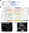
FIGURE 1.
FIGURE 1.
The ACCH domain defines a novel domain family encoded across metazoan genomes. A, the protein sequences of human Amot80 (NM_133265), Amot130 (130-kDa isoform of Amot; NM_001113490), JEAP/AmotL1 (NM_130847), and Mascot/AmotL2 (NM_016201) were aligned. The percentage identity between residues that collectively are predicted to form a coiled-coil fold (the region of highest identity between these proteins) and that are suggested here to compose the human ACCH domain members was computed by blast2seq (NCBI). B, putative proteins from the indicated metazoan genomes were predicted to encode ACCH domains based on their having a region of at least 200 residues that shared more conservation with human Amot ACCH than with any other region in any human protein. These proteins were iteratively aligned by blast2seq and by ClustalW alignments to human Amot. The conservation of residues between ACCH domains (ClustalW) are depicted at the left. The conservation between residues within the entire proteins with human Amot130 are depicted at the right (computed using Blast2seq/ClustalW alignments). A heat map indicates the percentage conservation. C, MDCK cells stably expressing YFP-tagged Amot80 were seeded on transwell filters, cultured for 2 days, and imaged live. A single confocal image is depicted. D, MDCK cells transiently transfected with a vector that expresses YFP AmotΔACCH were cultured and imaged. A single confocal image is depicted (top), and the y-z axis from reconstructed confocal sections from the region outlined in the red box is shown in the bottom image.
FIGURE 2.
FIGURE 2.
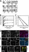
The ACCH domain selectively binds and penetrates membranes containing monophosphorylated phosphatidylinositols. A, the purified human Amot ACCH domain was incubated with liposomes of the indicated compositions (5% PIs or 20% PA, PS, PI, or PG). The supernatant (S) and pellet (P) following sedimentation was resolved by SDS-PAGE and visualized by Coomassie dye. B, the saturation responses (Req) (see supplemental Fig. S2B) at each ACCH domain protein concentration from SPR sensorgrams were plotted versus protein concentration for different liposomes (PM + PI(4)P (■), PM (□), POPC/POPE/PI4P (75:20:5) (▿), POPC/POPE/PI(3)P (75:20:5) (◇), POPC/POPE/PI(5)P (75:20:5) (♦), and POPC/POPE/PI(4,5)P2 (75:20:5) (▵)). The Kd was determined for a minimum of seven different protein concentrations by nonlinear least squares analysis of the binding isotherm using the equation, Req = Rmax/(1 + Kd/C). At least three replicates were done to calculate an S.D. value. C, insertion of the ACCH domain into POPC/POPE (80:20) (○), PM (□), POPC/POPE/PI(4)P (75:20:5) (▿), POPC/POPE/PI(3)P (75:20:5) (◇), and POPC/POPE/PI(4,5)P2 (75:20:5) (▵) monolayers as a function of π. 10 μg of ACCH was injected into the subphase of a monolayer of the indicated initial surface pressure. The Δπ was then monitored for 45 min to construct the curves and determine πc, the x intercept. D, MDCK cells stably expressing DsRed Amot80 and transiently expressing CFP-tagged FAPP1-PH were imaged live. The merge of the Amot (red) and FAPP1-PH (blue) is depicted on right. E, three-dimensional reconstructions of the x-y, x-z, and y-z dimensions of a live MDCK cell grown on a transwell filter expressing Amot ACCH (red) and CFP FAPP1-PH (blue). F, MDCK cells were transfected with vectors that express citrine Akt-PH (green, PIP3 probe), DsRed PLCδ PH (red, PIP2 probe) and cerulean Amot ACCH (blue) (left). Cells were then cultured on a transwell filter for 48 h and then imaged live. The x-z cut section from reconstructed confocal images is shown. mN, millinewtons.
FIGURE 3.
FIGURE 3.
The ACCH domain preferentially binds membranes enriched in cholesterol. A, relative response values (relative to POPC/POPE (80:20) control surface) were measured using SPR for 100 nm ACCH binding to POPC/POPE/cholesterol (80 − x:20:x) liposomes, where x equals the cholesterol concentration in each measurement. The S.D. value was calculated from five or more measurements for each respective cholesterol concentration. B, cellular membranes were prepared from MDCK cells expressing 3× FLAG-tagged Amot. Membranes were then separated by density using an Optiprep gradient, and the cholesterol content and the relative levels of Amot and caveolin levels in each fraction are depicted. The relative amounts of cholesterol were determined using an Amplex Red® kit (Sigma), whereas Amot and caveolin were detected by immunoblot analysis. C, fixed MDCK cells were permeabilized and immunostained for Amot (red) and for cholesterol as measured by binding to filipin (green) and with DAPI stain (blue). Confocal images of Amot (left), filipin (middle), and the merge showing DAPI stain (blue) (right) are shown. D, MDCK cells stably expressing DsRed Amot80 (red) were stained with filipin (green) and with DAPI stain (blue) and visualized as indicated. E, MDCK cells expressing DsRed Amot ACCH (red) were cultured on transwell filters with BODIPY cholesterol (green) and stained for nuclei with DAPI (blue). Live cells were then imaged as a single en face confocal section (main panel) and as x-y as well as x-z reconstructed z-stacks. F, MDCK cells stably expressing DsRed Amot80 were incubated with BODIPY ceramide and imaged live where a confocal section is depicted with DsRed Amot80 (left) and BODIPY ceramide (middle), and the merge is shown with Amot (red), BODIPY ceramide, and DAPI stain (blue) (right).
FIGURE 4.
FIGURE 4.
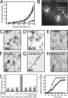
The ACCH domain selectively binds curved membranes and tubulates liposomes containing PI(4)P. A, liposomes with the indicated diameters were analyzed by SPR as described in the legend to Fig. 2 for binding of 100 nm Amot ACCH to POPC/POPE/PI(4)P (75:20:5) (▿), PM (□), POPC/POPE/POPS (60:20:20) (▴), and POPC/POPE/PI (60:20:20) (▾). Also, binding of 100 nm epsin ENTH was monitored to POPC/POPE/PI(4,5)P2 (75:20:5) (●) liposomes of the indicated diameters. B, MDCK cells that stably express DsRed Amot80 were imaged live at 2-s intervals for 80 s. A single frame from this sequence shows Amot marking highly dynamic and often tubulating endosomes (for the complete sequence, see supplemental Movie 2). C–H, liposome samples (1 mg/ml) prepared by extrusion through 100-nm membranes in POPC/POPE (80:20) (C), POPC/POPE/PI(4)P (75:20:5) (D), and POPC/POPE/PI(4,5)P2 (E) were applied to carbon-Formvar-coated copper grids (EMS), and membrane morphologies were examined on an FEI Magellan scanning electron microscope at an electron energy of 15 kV. Representative images were taken with a direct magnification of ×72,000–202,000. The scale bar in each panel is 400 nm. To assess the ability of the ACCH domain to induce lipid curvature changes, liposomes in POPC/POPE (80:20) (F), POPC/POPE/PI(4)P (75:20:5) (G), and POPC/POPE/PI(4,5)P2 (75:20:5) (H) were incubated with 5 μm ACCH domain for 15 min at 25 °C. The sample (8 μl) was then applied to the grids and negatively stained and imaged as described above. I, the size distributions of base POPC/POPE (80:20) liposomes with the indicated lipid ligand (POPC/POPE/PI(4)P (75:20:5) or POPC/POPE/PI(4,5)P2 (75:20:5) were prepared by 100-nm extrusions and assessed by dynamic light scattering analysis. Subsequently, liposomes of the indicated composition were incubated with the respective membrane-tubulating proteins. Reactions were carried to saturation, and then the average diameter of the membranes in each sample was determined by dynamic light scattering analysis. Each bar represents the mean diameter for the respective lipid. J, leakage of 5-carboxyfluorescein dye from liposomes of the indicated compositions by the addition of Amot ACCH protein (POPC/POPE (○), POPC/POPE/PI(4)P (▿), and POPC/POPE/PI(4,5)P2 (▵)) or ENTH domain (POPC/POPE/POPI(4,5)P2 (●)). Triton X-100 disruption of vesicles and subsequent leakage was used as a positive control (▴). At least three replicates were done to calculate an S.D. value for each time point.
FIGURE 5.
FIGURE 5.
Amot and Amot ACCH similarly distribute in MDCK cells at perinuclear endosomes with Rab11 and Arf6. A, MDCK cells were plated for 18 h, fixed with paraformaldehyde, and immunostained with a rabbit polyclonal antibody directed against Amot (left) and mouse monoclonal antibody directed against Rab-11 (middle) and DAPI stain to visualize nuclei. The merger of Amot (red), Rab11 (green), and DAPI stain (blue) is shown on the right. B, MDCK cells stably expressing DsRed Amot ACCH were plated for 24 h before being fixed with paraformaldehyde and immunostained for Rab11 and stained with DAPI. Left, DsRed Amot ACCH; middle, Rab11; right, merge of Amot (red), Rab11 (green), and DAPI (blue). C, MDCK cells were plated for 18 h and then immunostained for endogenous Amot (left) and Arf-6 (middle). The merger of Amot (red), Arf6 (green), and DAPI (blue) is shown on the right.
FIGURE 6.
FIGURE 6.
The ACCH domain promotes the interaction of Amot with polarity proteins at endosomal membranes. A, the relative association of Myc-tagged Amot80, AmotΔCOOH (which lacks 4 C-terminal residues), and AmotΔACCH (which lacks residues 1–242) with FLAG-tagged Mupp1 was examined. HEK 293T cells co-transfected with the indicated vectors were used for immunoprecipitations with anti-FLAG (M2) antibody. Immunoblots (IB) of lysates (left) and immunoprecipitants (IP) (right) using the indicated antibodies against FLAG (M2) and Myc (9E10) are shown. B, the relative association of FLAG-tagged Amot80 or AmotΔACCH with CFP-tagged Par-3 was examined. HEK 293T cells co-transfected as indicated were used for immunoprecipitation with anti-FLAG antibody. Immunoblots of lysates (left) and immunoprecipitants (right) with antibodies against FLAG and Par-3 are shown. C, a single en face confocal image of live MDCK cells showing the co-distribution of exogenously expressed DsRed Amot80 (left), YFP Patj (middle), and CFP Par-3 (right). MDCK cells were transfected with vectors expressing these three proteins and imaged live after removing calcium from the media. The movement of these proteins over 5 min is shown in supplemental Movies 3, A–C.

Similar articles

Cited by

References

    1. Nelson W. J. (2003) Nature 422, 766–774 - PMC - PubMed
    1. Nelson W. J. (2009) Cold Spring Harb. Perspect. Biol. 1, a000513. - PMC - PubMed
    1. Shin K., Fogg V. C., Margolis B. (2006) Annu. Rev. Cell Dev. Biol. 22, 207–235 - PubMed
    1. Kirby C., Kusch M., Kemphues K. (1990) Dev. Biol. 142, 203–215 - PubMed
    1. Tepass U., Knust E. (1993) Dev. Biol. 159, 311–326 - PubMed

Publication types

MeSH terms

LinkOut - more resources