. 2010 Mar 12;285(11):8148-54.
doi: 10.1074/jbc.M109.089821. Epub 2010 Jan 15.

Arginine methylation of vasa protein is conserved across phyla

Affiliations

Arginine methylation of vasa protein is conserved across phyla

Yohei Kirino et al. J Biol Chem. .

Abstract

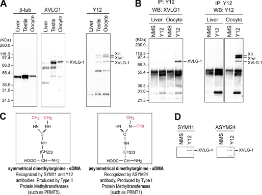
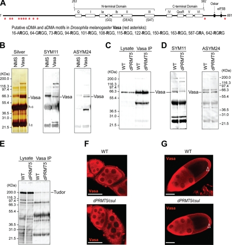
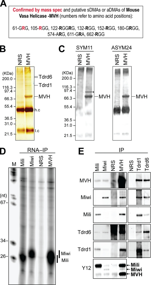
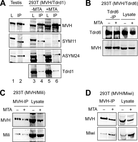
Recent studies have uncovered an unexpected relationship between factors that are essential for germline development in Drosophila melanogaster: the arginine protein methyltransferase 5 (dPRMT5/Csul/Dart5) and its cofactor Valois, methylate the Piwi family protein Aub, enabling it to bind Tudor. The RNA helicase Vasa is another essential protein in germline development. Here, we report that mouse (mouse Vasa homolog), Xenopus laevis, and D. melanogaster Vasa proteins contain both symmetrical and asymmetrical dimethylarginines. We find that dPRMT5 is required for the production of sDMAs of Vasa in vivo. Furthermore, we find that the mouse Vasa homolog associates with Tudor domain-containing proteins, Tdrd1 and Tdrd6, as well as the Piwi proteins, Mili and Miwi. Arginine methylation is thus emerging as a conserved and pivotal post-translational modification of proteins that is essential for germline development.


Figures

FIGURE 1.
FIGURE 1.
X. laevis Vasa protein XVLG1 is immunoprecipitated by Y12 and contains both sDMA and aDMA. A, Western blots from lysate of indicated X. laevis tissues. B, Y12 immunoprecipitates (IP) from X. laevis liver or oocytes were probed on Western blots (WB) with indicated antibodies (NMS: nonimmune mouse serum; negative control). C, arrangement of methyl groups in dimethylarginines. D, Y12 immunoprecipitates from X. laevis oocytes were probed on Western blots with anti-sDMA (SYM11) or anti-aDMA (ASYM24) antibodies. β-tub, β-tubulin.
FIGURE 2.
FIGURE 2.
D. melanogaster Vasa protein contains sDMAs and aDMAs, and Drosophila PRMT5 (dPRMT5/csul/dart5) is required for sDMA modifications. A, a schematic of Vasa and its putative DMAs is shown. The conserved DEAD-box helicase motifs and domains of Vasa that interact with other proteins (Oskar and eukaryotic initiation factor 5B (eIF5B) are also shown. B, Vasa or nonimmune mouse serum (NMS) immunoprecipitates (IP) from D. melanogaster ovaries were resolved by NuPAGE and stained by silver-staining or probed on Western blots with SYM11 or ASYM24 (NMS, nonimmune mouse serum; negative control). Vasa protein is indicated and was also confirmed by mass spectrometry (see supplemental Table 1). H.c, heavy antibody chain; l.c, light chain. Lysate or Vasa immunoprecipitates from WT or dPRMT5/csul ovaries were probed on Western blots with anti-Vasa (C); SYM11 and ASYM24 (D); and anti-Tudor (E). Localization of Vasa in WT and dPRMT5/csul ovaries in early (F) or late egg chambers (G); arrowhead indicates pole (germ) plasm. Scale bar is 40 μm.
FIGURE 3.
FIGURE 3.
Mouse Vasa protein MVH contains sDMAs and aDMAs and associates with Miwi, Mili, Tdrd1, and Tdrd6. A, confirmed and putative DMAs in MVH. Arg62 and Arg105 (red) were identified as dimethylated by mass spectrometry (mass spec) (see supplemental Table 3). B, MVH immunoprecipitates from mouse testis were resolved by NuPAGE and stained by silver-staining. Co-precipitating proteins (indicated) were identified by mass spectrometry (see supplemental Table 1). NRS, nonimmune rabbit serum (negative control). C, MVH immunoprecipitates were probed on Western blots with SYM11 or ASYM24; an asterisk indicates a protein band that likely corresponds to Miwi. D, RNA-immunoprecipitation with indicated antibodies from mouse testis. Miwi- and Mili-bound piRNAs are indicated. E, immunoprecipitates (IP; NRS, MVH, Miwi, Mili, Tdrd1, and Tdrd6) from mouse testis were probed on Western blots with indicated antibodies. nt, nucleotide; M, marker.
FIGURE 4.
FIGURE 4.
Association of MVH with Tdrd1, Tdrd6, Miwi and Mili in untreated and MTA-treated 293T cells. A, lysate (L) or MVH immunoprecipitates (IP) from mouse testis or 293T cells expressing MVH and Tdrd1, in the presence (+) or absence (−) of the methylation inhibitor MTA, were probed on Western blots with indicated antibodies. All filled circles indicate immunopurified MVH. Many methylated proteins are recognized by SYM11 and ASYM24 in the lysates of untreated 293T cells (lane 3 in A and B). Entire blot membranes for SYM11 and ASYM24 are shown in supplemental Fig. 2. B, Tdrd6 immunoprecipitates from 293T cells expressing MVH and Tdrd6, in the presence or absence of MTA, were probed on Western blots with anti-Tdrd6 or anti-MVH antibodies. MVH immunoprecipitates from 293T cells expressing MVH and either Mili (C) or Miwi (D) and in the presence (+) or absence (−) of MTA, were probed on Western blots with indicated antibodies.

Similar articles

Cited by

References

    1. Williamson A., Lehmann R. (1996) Annu. Rev. Cell Dev. Biol. 12, 365–391 - PubMed
    1. Strome S., Lehmann R. (2007) Science 316, 392–393 - PubMed
    1. Dansereau D. A., Lasko P. (2008) Methods Mol. Biol. 450, 3–26 - PMC - PubMed
    1. Rongo C., Lehmann R. (1996) Trends Genet. 12, 102–109 - PubMed
    1. Harris A. N., Macdonald P. M. (2001) Development 128, 2823–2832 - PubMed

Publication types

MeSH terms

Substances

LinkOut - more resources